Clear Sky Science · en
Charting spatial ligand-target activity using Renoir
How Cells Talk in Their Neighborhoods
Our bodies are made of countless cells that constantly "talk" to one another to keep tissues healthy, build organs, and, in some cases, drive diseases like cancer. This paper introduces Renoir, a computer method that reads large-scale gene activity maps to work out where and how this cellular chatter happens in real tissue. By combining modern single-cell and spatial genomics, Renoir helps researchers see not just who is talking to whom, but where in the tissue these conversations are strongest and what effects they have.
Signals, Messengers, and Cellular Conversations
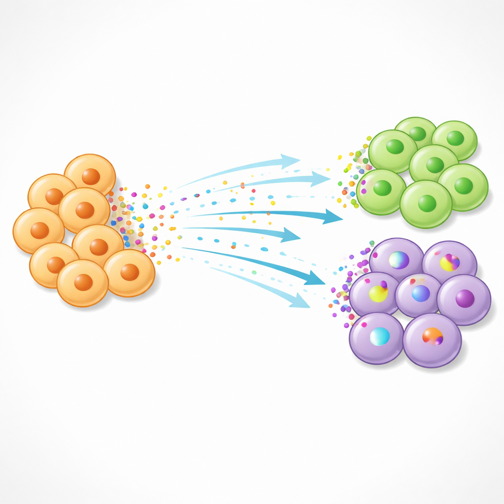
Cells communicate using small protein messengers called ligands that are released by one cell and sensed by neighboring cells through receptors on their surface. When a ligand binds its receptor, it can switch on a cascade of “target” genes inside the receiving cell, changing its behavior. Many existing tools try to infer these interactions from gene activity data, but they often ignore where cells are physically located. Because many signals act only over short distances, losing this spatial context can lead to misleading results—showing apparent communication between cell types that in reality sit far apart in the tissue.
What Makes Renoir Different
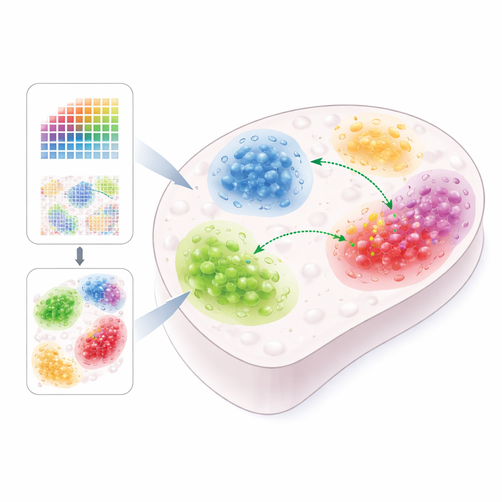
Renoir is designed specifically to bring space back into the picture. It takes in either single-cell–resolution spatial datasets or a combination of lower-resolution spatial data and classic single-cell data from the same tissue. Using curated lists of ligands and their potential target genes, Renoir calculates a “neighborhood activity score” for each ligand–target pair at each location in the tissue. This score blends several pieces of information: which cell types are present nearby, how strongly they express the ligand and the target gene, whether the receiver cells actually express the right receptor, and how tightly ligand and target tend to vary together across cell types. The result is a spatial map that highlights where particular signaling relationships are likely to be active.
Finding Hidden Neighborhoods in Healthy and Diseased Tissues
Once neighborhood scores are calculated, Renoir can group locations into “communication domains”—patches of tissue that share similar patterns of signaling. Applied to mouse brain data, these domains lined up with known brain regions and revealed region-specific communication between astrocytes and different types of neurons. In triple-negative breast cancer, Renoir uncovered distinct tumor niches in which cancer cells, immune cells, and connective tissue cells exchange signals linked to growth, invasion, and immune suppression. In developing human fetal liver, it identified a niche where liver cells (hepatocytes) and specialized macrophages interact through a molecule called plasminogen, pointing to a role in liver growth and remodeling.
Testing Renoir Against Other Methods
The authors rigorously tested Renoir by creating semi-synthetic datasets where the true signaling patterns were known in advance. They compared Renoir to several leading tools that infer cell–cell communication from spatial data. Across tissues such as intestine, brain, and breast cancer, Renoir more accurately distinguished locations with real ligand–target activity from those without, and it was less likely to report false interactions in places lacking the right receptors. Even when the data were made noisier—by reducing sequencing depth or scrambling some cell labels—Renoir’s performance remained stable. In a well-studied region of the human brain, the method’s inferred communication domains matched expert-defined tissue layers better than competing approaches.
From Maps of Communication to Therapeutic Clues
Renoir is not just a mapping tool; it can also rank which ligands are most influential in each domain and summarize the activity of whole signaling pathways. In liver cancer, this highlighted “onco-fetal” signaling circuits where tumor-associated cells reuse developmental programs seen in fetal liver. Renoir predicted that ligands such as interleukin-6 from this niche could reprogram nearby stem-like liver cells; laboratory experiments confirmed that interleukin-6 pushes liver cancer cells toward a more stem-like state. Altogether, the study shows how combining spatial genomics with smart computation can turn static gene maps into dynamic portraits of cellular dialogue, offering new entry points for therapies that aim to disrupt harmful conversations while preserving healthy ones.
Citation: Rao, N., Kumar, T., Kazemi, D. et al. Charting spatial ligand-target activity using Renoir. Nat Commun 17, 3983 (2026). https://doi.org/10.1038/s41467-026-72388-7
Keywords: spatial transcriptomics, cell communication, ligand signaling, tumor microenvironment, computational biology
See more on the researcher's website: https://sites.google.com/view/cosmiclab-iitk/home