Clear Sky Science · en
PARP1-HPF1 structure and dynamics on nicked DNA suggest a mechanism for acute and localized ADP-ribosylation
How cells sense tiny breaks in their DNA
Every cell in your body is constantly bombarded by events that nick or break its DNA. If these lesions are not detected and fixed quickly, they can lead to mutations and, ultimately, cancer. This study reveals how one of the cell’s key first responders, a protein called PARP1 working together with a partner protein HPF1, recognizes a tiny break in DNA and then rapidly decorates nearby proteins with chemical tags. These tags act like a bright flare, summoning and organizing the repair crew right where it is needed.
A molecular first responder at work
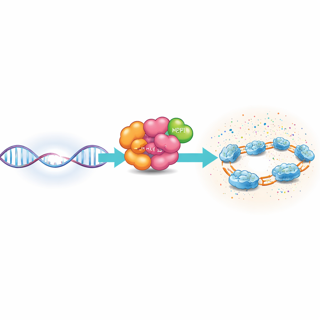
PARP1 is an abundant guardian protein that scans DNA for damage. When it encounters a single-strand nick – a cut in just one of the two DNA strands – PARP1 docks onto the break using several specialized regions that grip the DNA. Once activated, PARP1 uses a cellular fuel molecule, NAD+, to build chains of ADP-ribose on itself and on nearby proteins, especially histones, which help package DNA into chromatin. These modifications briefly loosen the local chromatin structure and attract repair factors, creating a tightly focused repair zone around the damaged site.
Seeing the full machine on broken DNA
Until now, most structural snapshots of PARP1 had shown only parts of the protein, leaving open the question of how the full-length molecule assembles on a DNA break together with its partners. The authors used single-particle cryo–electron microscopy, a technique that images frozen molecules at near-atomic detail, to visualize full-length PARP1 bound to a nicked piece of DNA along with HPF1 and a fragment of another partner protein called Timeless. They combined these views with single-molecule fluorescence experiments and solution scattering measurements to understand not only the structure, but also the motion, of this complex in action.
Bending DNA and organizing the scaffold
The images reveal that several PARP1 regions clamp around the nick and sharply bend the DNA by about 75 degrees. Three zinc-finger segments and a WGR domain form a loose but coordinated scaffold that anchors PARP1 directly at the break. An adjoining region called the helical domain joins this assembly, helping to stabilize the DNA-bound state. Together, these pieces create a rigid anchor at the damage site, while other parts of PARP1 – notably a BRCT domain closer to the middle of the protein – remain flexible and often invisible in the images, indicating they move freely even when the DNA-binding core is locked in place.
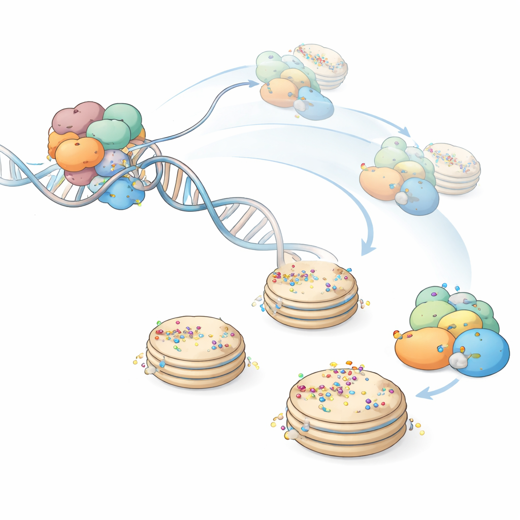
A catalytic arm on a flexible leash
The most striking discovery is what happens to PARP1’s catalytic region, the part that actually builds ADP-ribose chains. In earlier crystal structures, this catalytic block sat snugly against the helical domain in an “off” state that blocked access to NAD+. In the new cryo-EM views, once PARP1 is fully organized on nicked DNA, this catalytic block largely detaches from its helical neighbor and becomes highly mobile, remaining connected only by flexible linkers. HPF1 binds directly to this mobile catalytic region, reshaping its active site so that serine residues on histones become preferred targets. Single-molecule fluorescence measurements confirm that binding of a NAD+-mimicking compound both stabilizes PARP1 on DNA and reduces DNA bending fluctuations, consistent with an activated yet dynamically tethered catalytic arm.
A local but powerful chemical flare
By combining structural imaging, single-molecule dynamics, and solution scattering, the authors propose a model in which binding to a DNA nick organizes the N-terminal half of PARP1 into a rigid clamp while freeing the catalytic region at the other end to swing around on a “leash.” This mobile yet constantly active catalytic arm, in partnership with HPF1, can rapidly tag PARP1 itself and nearby histones with ADP-ribose chains within a limited radius surrounding the break. The result is an acute but highly localized burst of signaling that remodels chromatin and recruits repair factors precisely where they are needed, helping cells maintain genome stability while minimizing unnecessary disruption elsewhere in the nucleus.
Citation: Sverzhinsky, A., Xue, H., Langelier, MF. et al. PARP1-HPF1 structure and dynamics on nicked DNA suggest a mechanism for acute and localized ADP-ribosylation. Nat Commun 17, 2825 (2026). https://doi.org/10.1038/s41467-026-69375-3
Keywords: DNA repair, PARP1, ADP-ribosylation, cryo-EM, chromatin