Clear Sky Science · en
TREM2-mediated microglial phagocytosis of inhibitory synapses contributes to prolonged FS-induced epileptogenesis
Why childhood fevers can leave a lasting mark on the brain
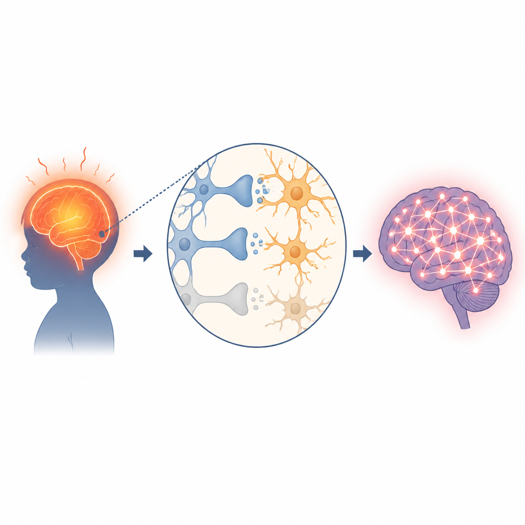
Many parents are told that febrile seizures, the convulsions that sometimes accompany high fevers in young children, are usually harmless. Yet some children later develop epilepsy, a condition marked by repeated unprovoked seizures. This study in young rats explores what may be happening inside the brain during long-lasting febrile seizures and points to a specific immune-related switch on brain cells that could help explain how a brief illness leaves a long-term electrical scar.
Brain cells that clean up connections
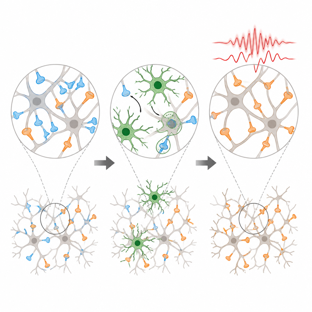
The brain is wired by trillions of tiny junctions called synapses, where nerve cells pass signals to one another. A healthy brain keeps a careful balance between “go” signals that excite activity and “stop” signals that calm things down. Special immune cells in the brain, called microglia, act as housekeepers: they patrol this wiring and nibble away at synapses that are weak, damaged, or no longer needed. This pruning helps shape brain circuits during development and in response to experience.
A molecular handle for pruning
Microglia rely on surface receptors to decide when and where to prune. One such receptor, TREM2, helps microglia recognize synapses marked for removal. When TREM2 levels rise, microglia become more active and more eager to engulf synapses. In the rat model used in this study, long-lasting febrile seizures in early life sharply boosted TREM2 in key brain regions that help control seizures. At the same time, the density of calming, inhibitory synapses dropped, while the number of stimulating, excitatory synapses rose, tilting brain circuits toward overactivity.
Turning the pruning dial up and down
The researchers then tested ways to nudge this system. Lowering TREM2 directly with a genetic tool, or indirectly by activating another receptor called CD33, quieted microglia and reduced their digestion of inhibitory synapses. In these rats, more calming synapses were preserved, and it took a higher dose of a seizure-triggering drug to provoke strong seizures. Their brain wave recordings also showed fewer and shorter seizure episodes. These results suggest that dampening TREM2 activity can protect the balance of synapses and make the brain less seizure-prone after prolonged febrile events.
When “eat me” signals are blocked
The team also asked what happens if microglia are prevented from seeing one of their main clues that a synapse needs removal. A fat molecule called phosphatidylserine normally flips to the outer surface of stressed synapses and acts as an “eat me” flag for TREM2. The researchers used a protein, annexin V, to cover this molecule so TREM2 could not recognize it. As expected, inhibitory synapses were less likely to be engulfed. Surprisingly, however, seizures became worse, and brain activity grew more chaotic. Damage signals and markers of oxidative stress and inflammation rose sharply, and other support cells called astrocytes became overactive and promoted new excitatory synapses.
How this work reshapes our view of febrile seizures
These findings paint a more nuanced picture of the brain’s cleanup crew. After prolonged febrile seizures, TREM2-driven microglia appear to over-prune calming synapses, leaving circuits easier to trigger into epilepsy. Gently lowering TREM2 can restore some balance and reduce seizure risk in this animal model. But completely blocking the signals that guide microglia to damaged sites may backfire, allowing harmful debris and inflammation to build up and driving other pathways that strengthen excitatory wiring. For lay readers, the take-home message is that the way the brain’s own immune cells respond to early-life fevers may help determine whether a child’s brief convulsion remains an isolated event or contributes to a chronic seizure disorder later on.
Citation: Wang, X., Zhou, H., Zhai, Y. et al. TREM2-mediated microglial phagocytosis of inhibitory synapses contributes to prolonged FS-induced epileptogenesis. Cell Death Discov. 12, 223 (2026). https://doi.org/10.1038/s41420-026-03118-7
Keywords: febrile seizures, epilepsy, microglia, TREM2, synaptic pruning