Clear Sky Science · en
Correlation networks of blood proteins in the neuroimmunology of schizophrenia—replication and extension
Why blood can hint at future mental illness
Schizophrenia often appears in late adolescence or early adulthood, and doctors still cannot reliably tell who will go on to develop a full psychotic disorder. This study looks for early warning signs not in brain scans, but in patterns among proteins floating in the blood. By examining how certain blood proteins rise and fall together in young people at high risk, the researchers hope to glimpse changes tied to brain wiring, blood clotting, and inflammation long before illness fully takes hold.
Following young people on the edge of psychosis
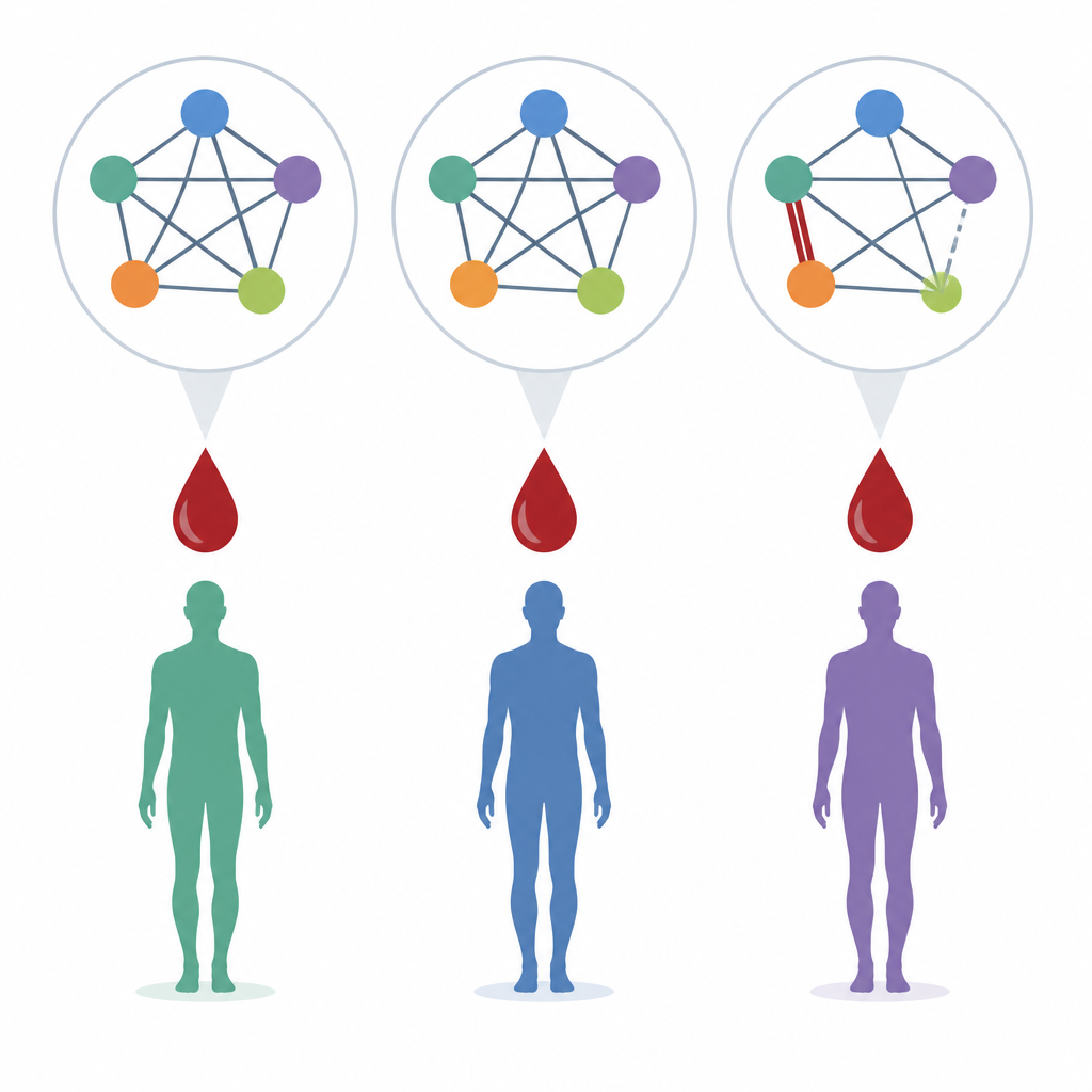
The work builds on two large North American projects that follow teenagers and young adults who show subtle, early warning symptoms of psychosis. These individuals are labeled clinical high risk and have about a one-in-five chance of developing a clear psychotic disorder within two years. In both study waves, called NAPLS2 and NAPLS3, researchers collected blood samples at the start, then tracked who later converted to psychosis, who stayed symptomatic but stable, and who were unaffected community volunteers. Rather than focus only on whether single proteins were higher or lower, the team examined whether pairs of proteins moved together more tightly in some groups than in others.
Two proteins that move in lockstep
Earlier work in NAPLS2 had highlighted a pair of blood proteins, SERPINE1 and TIMP1, that showed unusually strong coordination in people who later developed psychosis compared with those who did not. Both proteins are involved in slowing the breakdown of blood clots and in limiting the remodeling of the tissue scaffold that supports cells, including brain cells. In the new and larger NAPLS3 group, the same pattern appeared again: the correlation between SERPINE1 and TIMP1 was clearly higher in converters than in nonconverters or community volunteers. Sophisticated statistical checks, including permutation tests that shuffle the data thousands of times, suggested that seeing such similar patterns in both cohorts would be unlikely to occur by chance.
Clues from blood clotting and brain scaffolding
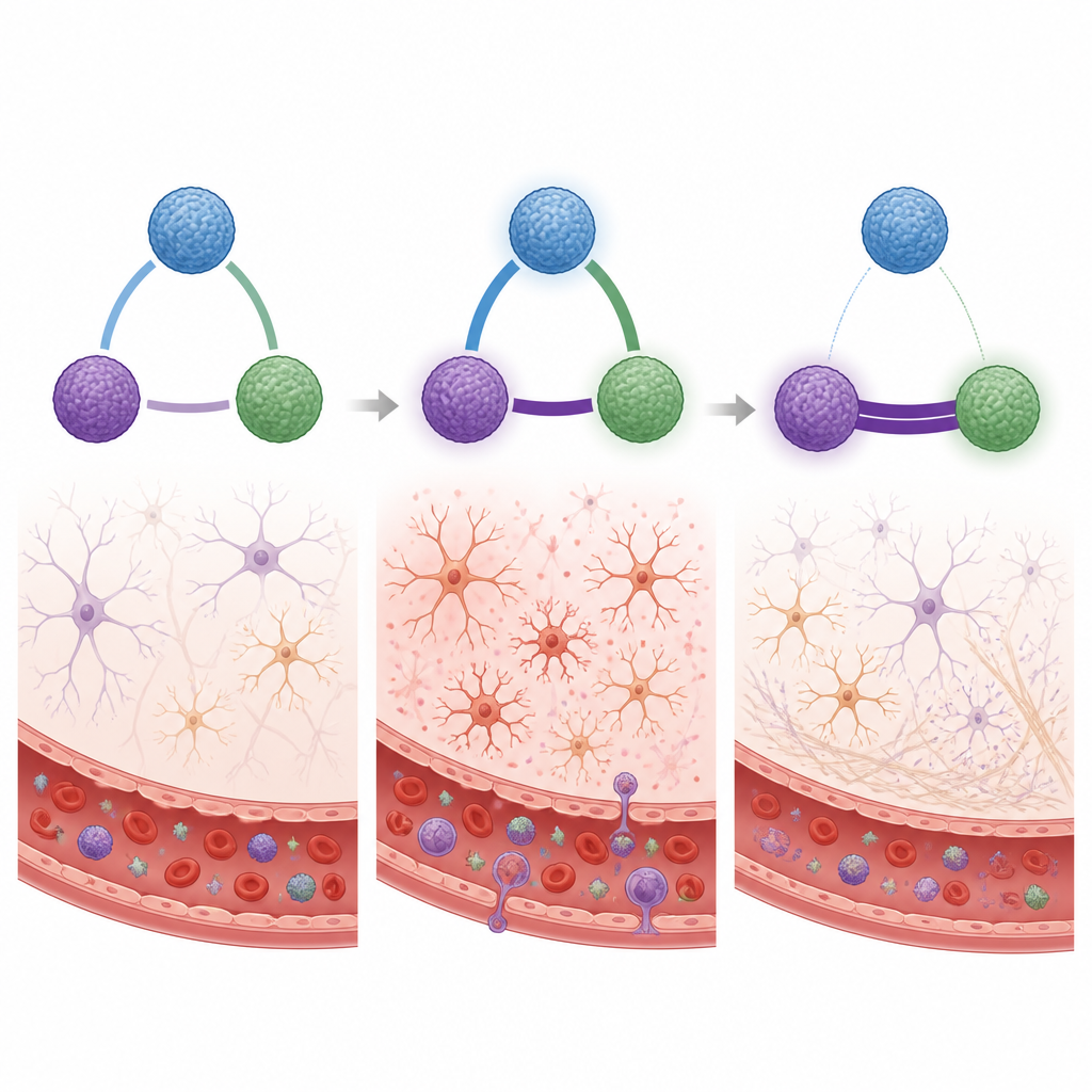
The team then added two more proteins to the picture, PLAT and PLAU, which help dissolve clots and reshape tissue, and are normally held in check by SERPINE1. In the new data, the link between PLAT and SERPINE1 was weaker in converters than in nonconverters, and the link between PLAU and SERPINE1 in converters actually tended to be negative. These shifts hint that the fine balance between forming and dissolving clots may be disturbed in those who progress to psychosis. At the same time, the strong partnership between SERPINE1 and TIMP1 suggests a system tilted toward preserving the existing tissue scaffold instead of allowing flexible reshaping of brain circuits. This ties in with other research showing abnormal loss of gray matter and changes in the specialized mesh, called perineuronal nets, that wraps certain brain cells during key learning windows.
How protein networks may reflect brain change
To better understand how these proteins fit together, the authors used existing databases of protein interactions. These maps show SERPINE1, TIMP1, PLAT, and PLAU as part of a wider web that governs clotting, the integrity of blood vessels, and the structure around neurons. Signals such as the molecule TGFB1 can spur cells to secrete both SERPINE1 and TIMP1, potentially explaining why their blood levels become tightly coupled when certain pathways are activated. Other studies have linked these same proteins to changes in the blood–brain barrier, to responses to psychedelic drugs that briefly reopen learning windows, and to the action of antipsychotic medications in cell models. Together, these lines of evidence suggest that altered blood protein relationships may mirror shifts in how the brain maintains and remodels its wiring.
What this might mean for future care
The findings do not yet offer a simple blood test that can predict schizophrenia in an individual person, and the authors stress that more data and better mathematical tools are needed. Still, the repeated observation that SERPINE1 and TIMP1 move more closely together in those who convert to psychosis points to biological systems worth watching. It hints that disturbed control of clotting and of the brain’s supporting matrix may be part of how psychosis develops. In the long run, tracking such protein networks could help researchers identify who is most at risk and design treatments that gently shift these systems back toward healthier patterns.
Citation: Jeffries, C.D., Bizon, C.A., Ford, J.R. et al. Correlation networks of blood proteins in the neuroimmunology of schizophrenia—replication and extension. Transl Psychiatry 16, 251 (2026). https://doi.org/10.1038/s41398-026-03934-6
Keywords: schizophrenia risk, blood proteins, extracellular matrix, coagulation, psychosis biomarkers