Clear Sky Science · en
A transcriptional program associated with neurotransmission in the living human brain
Why This Living Brain Study Matters
Most of what we know about the human brain’s molecules comes from tissue studied after death, long after electrical signaling has stopped. This paper turns that approach on its head. By briefly sampling brain tissue during routine surgery and recording live electrical activity at the same time, the researchers identify a coordinated set of genes that seem to support the very act of brain cells talking to each other. Understanding this “operating system” for brain communication could eventually reshape how we think about cognition, mental illness, and new treatments.
Peeking Inside the Working Human Brain
The team worked within the Living Brain Project, which collaborates with patients undergoing deep brain stimulation surgery for conditions such as Parkinson’s disease. During these operations, surgeons can safely remove a tiny sample from the front of the brain, an area involved in planning, decision making, and emotion. At nearly the same moment, thin recording electrodes measure activity in deeper structures that talk to this frontal region. In a subset of surgeries, patients even played a simple bargaining computer game while signals from brain chemicals like dopamine and serotonin were captured in real time. These paired tissue samples and recordings allowed the scientists to ask a rare question: which genes are turned up or down in living human brain cells when communication between regions is actually happening? 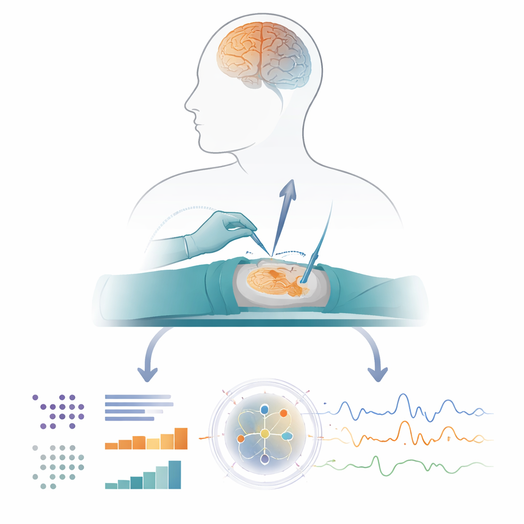
Finding Patterns in a Sea of Genes
Because each tissue sample contains thousands of genes, the researchers treated the data like a massive puzzle. They applied standard statistical tools to see whether small changes in electrical or chemical signals—such as fluctuations in dopamine during the bargaining game, or broad rhythm changes in deeper nuclei—were consistently linked to changes in gene activity in various cell types. Single-cell methods revealed how different brain cells (excitatory and inhibitory neurons, support cells like astrocytes and oligodendrocytes, and immune-like microglia) each carried their own molecular fingerprints. Even though the number of patients in some experiments was modest, the authors detected broad transcriptome-wide “signatures”: patterns across many genes that shifted together with live measures of neurotransmission.
Building a Shared Molecular Program
To check that these patterns were not statistical flukes, the team repeated the logic in independent data. One dataset came from other patients in the Living Brain Project where a different kind of recording, called microelectrode recordings, captured the balance of excitation and inhibition in deep nuclei. Another came from a published epilepsy study in which brain rhythms were recorded from patients’ temporal lobes before that tissue was surgically removed. Across these very different settings—different recording technologies, brain regions, and patient groups—the same groups of genes kept reappearing. The authors then used network-style analysis to find a core set of 588 genes that showed consistent associations with neurotransmission across at least two of the three independent experimental designs. They termed this shared set the “transcriptional program associated with neurotransmission,” or TPAWN. 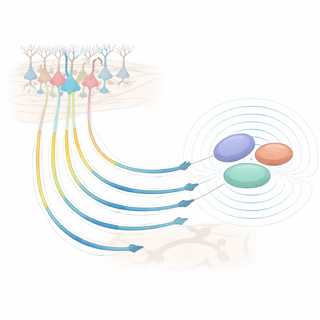
Linking Gene Programs, Circuits, and Disease
Once TPAWN was defined, the researchers asked what it might actually do. They found that these genes were enriched for roles in classic brain communication pathways, including synapses, ion channels, and long-term changes in connection strength. Compared with other brain-expressed genes, TPAWN genes were also more “evolutionarily constrained,” meaning harmful changes in them are rarely seen in large human populations—usually a sign that they are crucial for survival or healthy function. In a large New York City health system, people carrying rare disruptive variants in TPAWN genes had a higher risk of medical records reflecting hallucinations, hinting at a link between this program and mental health. At the cellular level, in the frontal cortex of living patients, cells with higher TPAWN activity looked most like a subtype of excitatory neurons that send long-range projections down to deep brain structures, matching the very circuits being recorded during surgery.
What This Means for Understanding the Brain
For a non-specialist, the key takeaway is that the brain’s electrical chatter is not just random sparks; it is tightly coordinated with a deeply conserved gene program that keeps communication channels tuned. This study provides the first robust map of that program directly in living human brain tissue, rather than in animal models or postmortem samples. While far from yielding new drugs tomorrow, the work lays a foundation: by tying together gene activity, cell types, brain circuits, and real-time behavior, it points toward molecular targets that may underlie cognition and psychiatric symptoms. Future, larger studies using similar live-tissue approaches could refine this program and eventually guide therapies that adjust brain circuits at their molecular roots.
Citation: Charney, A.W., Liharska, L.E., Vornholt, E. et al. A transcriptional program associated with neurotransmission in the living human brain. Mol Psychiatry 31, 2727–2738 (2026). https://doi.org/10.1038/s41380-025-03420-3
Keywords: neurotransmission, gene expression, prefrontal cortex, deep brain stimulation, brain circuits