Clear Sky Science · en
Mitochondrial hyperoxidation contributes to warm ischemia-reperfusion injury in rat and pig livers
Why this matters for donated livers
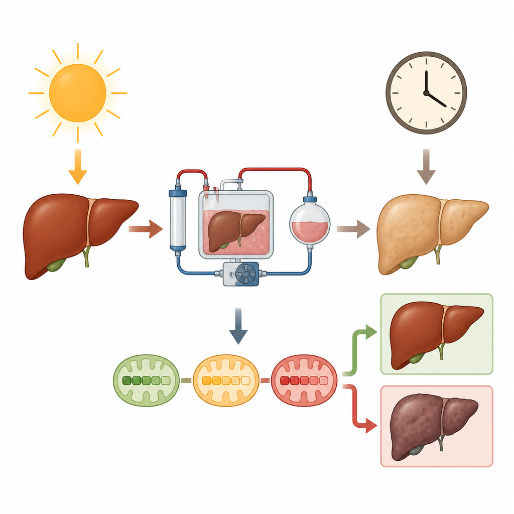
Many people die waiting for a liver transplant because too few donated organs are considered safe to use. One big reason is damage that occurs when blood flow stops and then restarts, a process called warm ischemia-reperfusion injury. This study explores a light-based way to watch tiny power plants inside liver cells in real time and tests a common blue dye as a potential way to protect these organs, with the goal of making more livers suitable for transplant.
Watching cell power plants with light
At the heart of this work are mitochondria, the parts of cells that turn oxygen and nutrients into usable energy. When their delicate machinery goes wrong, cells can fail and die. Existing methods to check mitochondrial health usually require taking tissue samples and complex lab processing, which is slow and impractical during organ preservation. The researchers built a resonance Raman spectroscopy system, which shines a gentle laser on the liver surface and reads back a spectral fingerprint from special light-absorbing groups inside mitochondrial proteins. From these fingerprints, they can estimate how oxidized or reduced different parts of the energy chain are without cutting into the organ.
Modeling damage in rat livers
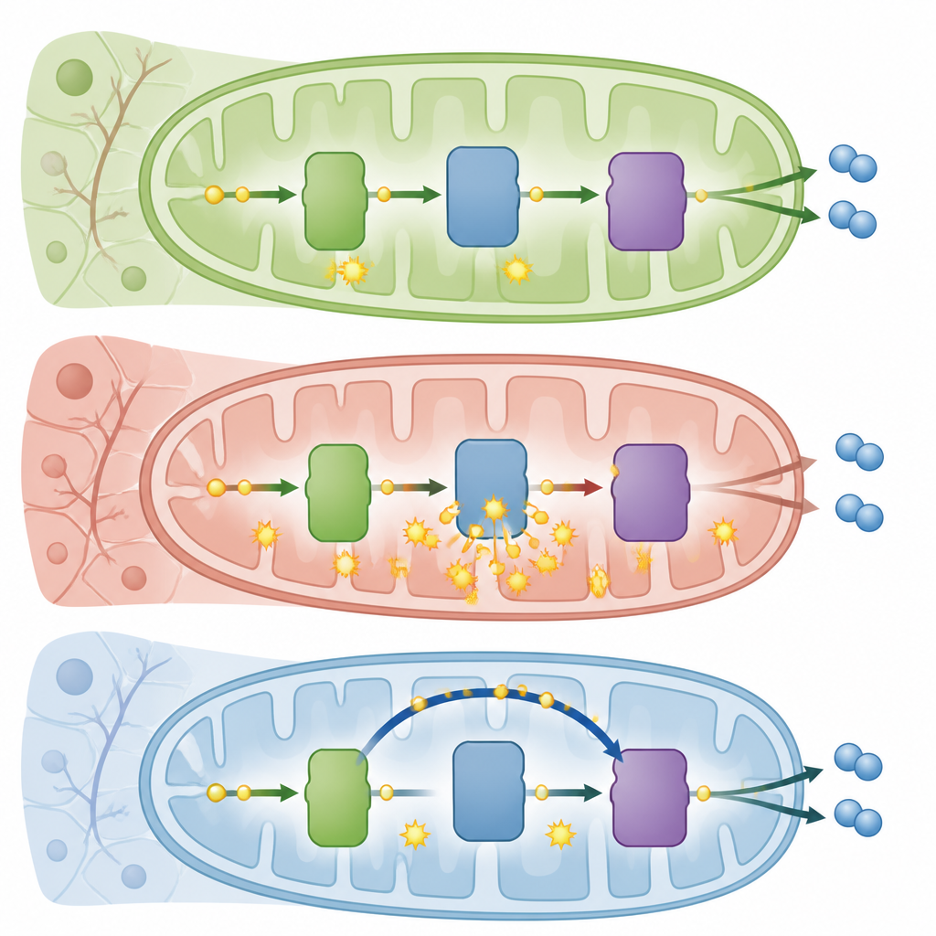
To understand how warm ischemia harms livers, the team used rat organs exposed to either no injury, one hour of warm blood stoppage, or a severe three-hour pause before machine perfusion at cool room temperature. They tracked not only standard measures like blood flow, oxygen use, and leak of injury enzymes, but also mitochondrial redox state across the whole organ and within two key steps of the energy chain, called complexes III and IV. The sickest livers, with three hours of warm ischemia, looked pale, showed poor blood flow and energy stores, and released more cell death markers. At the mitochondrial level, these organs showed an unusually hyperoxidized pattern, especially at complex III, suggesting electrons were leaking away instead of being passed down the chain to make energy.
Stress tests and a blue dye workaround
To probe this damage more deeply, the researchers briefly cut off oxygen again during perfusion and watched how rapidly mitochondria became reduced. Healthy and mildly injured livers responded quickly, while the severely injured livers changed much more slowly, consistent with a leaky, hyperoxidized system. The team then tested methylene blue, a long-used medical dye that can redirect electrons around complex III straight to the next step in the chain. In rat livers with severe warm ischemia, adding methylene blue shifted complex III to a fully oxidized state and increased the reduced fraction at complex IV, indicating that electrons were successfully bypassing the damaged region. These changes were accompanied by better oxygen use, lower lactate levels, and improved energy balance, especially when combined with an added oxygen carrier in the perfusion fluid.
Scaling up to pig livers like those used in clinics
Because pig livers resemble human livers in size and structure, the team next applied their strategy to pigs whose organs were recovered after controlled circulatory death. Some livers experienced a typical borderline duration of warm ischemia, while others endured a longer, normally disqualifying period. Using the same perfusion setup, they found that methylene blue, with or without the extra oxygen carrier, improved blood flow and boosted oxygen consumption in injured pig livers. Remarkably, livers that had suffered extended warm ischemia but received methylene blue alone reached hemodynamic and oxygen–lactate profiles similar to those of fresh, brain-death donor controls, even though some molecular injury markers still differed.
What this could mean for future transplants
Overall, the study shows that noninvasive light measurements can reveal a distinct signature of mitochondrial hyperoxidation at complex III during warm ischemia-reperfusion injury and that methylene blue can functionally bypass this damaged step to restore more normal energy use in rat and pig livers. For a layperson, the takeaway is that doctors may one day be able to “look” at an organ’s microscopic power system in real time and apply targeted treatments while the liver is on a machine, potentially rescuing organs that would otherwise be discarded. If confirmed in further work and eventually in humans, this approach could help expand the pool of usable donor livers and deepen our understanding of how oxygen-related injury unfolds in many diseases.
Citation: Nguyen, K.T., Ozgur, O.S., Jain, R. et al. Mitochondrial hyperoxidation contributes to warm ischemia-reperfusion injury in rat and pig livers. Commun Med 6, 307 (2026). https://doi.org/10.1038/s43856-026-01551-4
Keywords: liver transplantation, mitochondria, ischemia reperfusion, resonance Raman, methylene blue