Clear Sky Science · en
Single-scan detection of ligand-binding using hyperpolarization and low-field relaxation
Why this matters for future medicines
Designing new drugs often starts with a simple question: does this tiny molecule actually stick to the protein we care about? Nuclear magnetic resonance (NMR) is a powerful way to watch such binding in solution, but it normally needs large amounts of valuable protein. This study introduces a clever way to shrink those requirements by supercharging the NMR signal and exploiting how molecules move at low magnetic field, opening the door to more efficient and economical drug discovery experiments.
Watching tiny magnets inside molecules
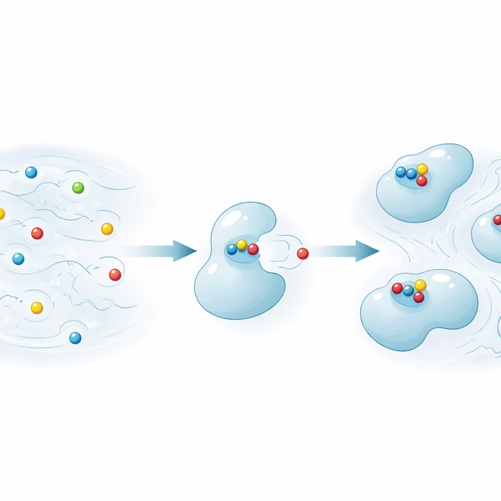
NMR works by detecting the behavior of atomic “spins,” tiny magnets inside nuclei such as hydrogen or carbon. When a small molecule ligand binds to a big protein, its motion slows down, and this affects how quickly its spins relax back to their resting state after being excited. Traditional NMR screening methods mainly track sideways relaxation (called T2), which changes when a ligand binds. In contrast, the authors focus on longitudinal relaxation (T1), which also reacts strongly to how fast a molecule tumbles but becomes much more informative at lower magnetic fields than those usually used for high-resolution NMR.
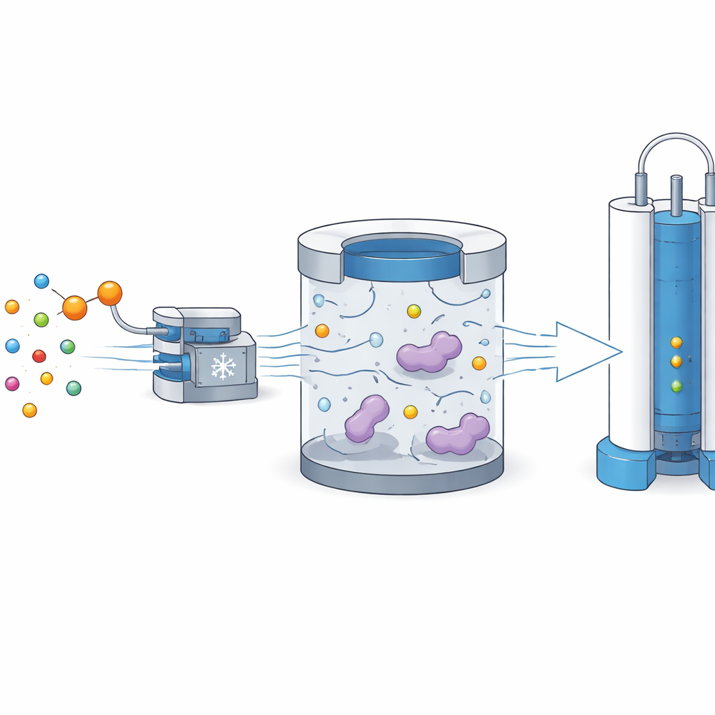
Boosting signals and using low-field waiting time
The team combines two ideas: hyperpolarization and low-field relaxation. First, they dramatically boost the signal of a specially labeled carbon atom in a reporter ligand using a method called dynamic nuclear polarization at very low temperature and high field. Then, instead of measuring immediately, they quickly dissolve the sample and move it into a moderate magnetic field of about 1.3 tesla, where the ligand is mixed with the target protein. During a 10-second waiting period at this lower field, ligands that bind the protein relax much faster than those that stay free, because their motion has slowed and they experience stronger fluctuating magnetic fields.
Reading out binding in a single NMR shot
After this low-field pause, the solution is transferred into a conventional high-field NMR magnet. There, the remaining enhanced carbon polarization of the ligand is converted into detectable hydrogen signals using a standard pulse sequence. The researchers record two spectra from each hyperpolarized shot: the first mainly reflects how much T1 relaxation occurred at low field, while the second probes a T2-like process under a spin-lock. By comparing the intensities of these two spectra with and without protein, they define simple scores that report whether binding has happened. Using just 14 micromolar of a carbon-13 labeled pyruvate reporter, they can clearly detect binding to a cancer-related enzyme, PHD1, even when the protein concentration is only 2 micromolar—and they do so in a single scan instead of many repeated measurements.
Testing competition and sensitivity
The method is also able to reveal when an unlabeled ligand competes for the same binding site. The authors add a strong competitor molecule, which displaces the labeled reporter from the protein. As the competitor blocks the protein from interacting with the reporter, the reporter’s signal at high field rebounds toward its protein-free level. This change shows up most clearly in the T1-based low-field score, while the more conventional T2-based measure sometimes stays near the noise level. Repeating experiments under similar conditions shows that the hyperpolarization process is repeatable enough that changes in signal due to protein binding—and its suppression by a competitor—stand out reliably.
What this means for drug discovery
In simple terms, the authors have turned the low-field waiting time into a highly sensitive test of whether a molecule binds to a protein. By starting with a hyperpolarized ligand, they can use very low concentrations but still see strong NMR signals, and by measuring how much signal is lost during the low-field pause, they obtain a clear contrast between bound and unbound states. This approach cuts the needed protein concentration down to the low micromolar range, a major advantage when proteins are hard to produce. With further refinements, such as doing more of the experiment at low field or in microfluidic devices, this strategy could become a practical tool for screening drug candidates while consuming far less precious protein material.
Citation: Narwal, P., Lorz, N., Minaei, M. et al. Single-scan detection of ligand-binding using hyperpolarization and low-field relaxation. Commun Chem 9, 140 (2026). https://doi.org/10.1038/s42004-026-01934-7
Keywords: NMR ligand binding, hyperpolarization, low-field relaxation, drug discovery, protein–ligand interactions