Clear Sky Science · en
Orbitofrontal PV interneurons modulate social interaction via default mode network dynamics
Why Our Brains Need Other People
Most of us feel that spending time with friends or family is good for our mental health. When this sense of connection fades, as in depression, schizophrenia, or Alzheimer’s disease, people often withdraw from others. This paper explores a key brain circuit that helps support social life. By studying mice, the researchers show how a small group of nerve cells in the front of the brain can disturb a broader “at-rest” brain network and, in turn, reduce normal social interaction and memory.
A Quiet Brain Network with a Big Job
Even when we sit still and let our minds wander, certain brain regions hum together in a coordinated pattern known as the default mode network, or DMN. In people, this network is linked to daydreaming, self-reflection, remembering the past, and thinking about others. Clinical studies from the PRISM project have found that people with schizophrenia or Alzheimer’s disease often show weakened connections inside this network, and those changes track with how socially withdrawn they are. The new study asks a causal question: if scientists deliberately disrupt DMN-like connectivity in an animal, does social behavior suffer?
A Small Control Hub in the Frontal Brain
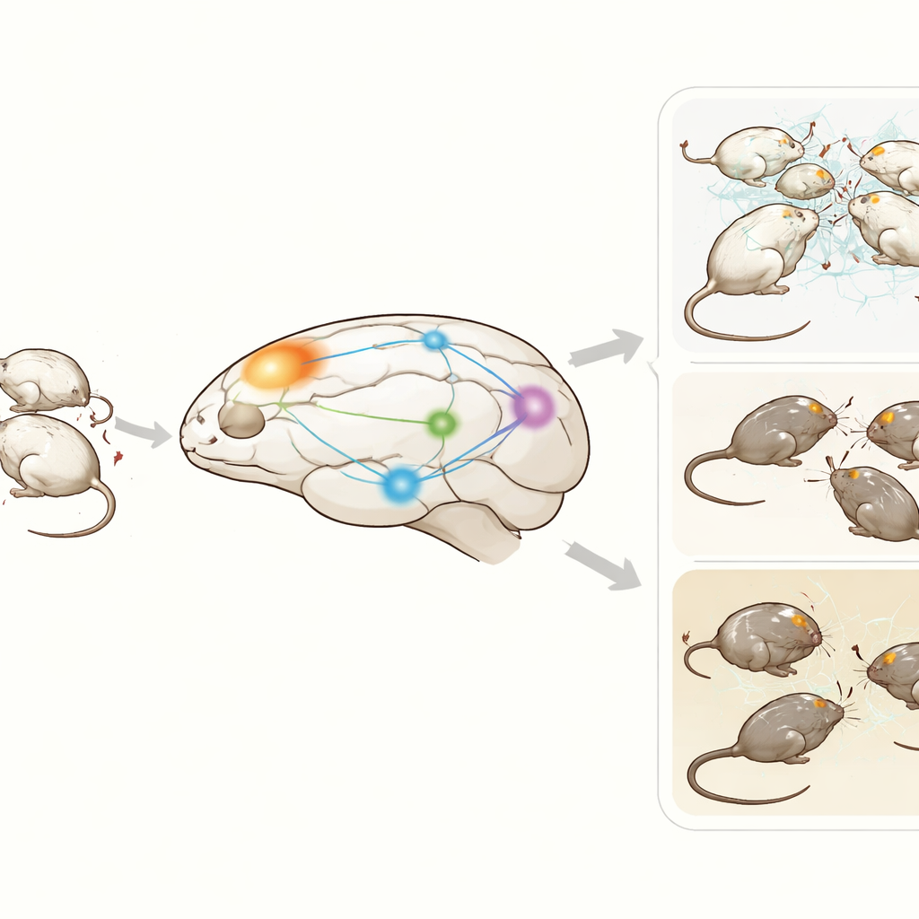
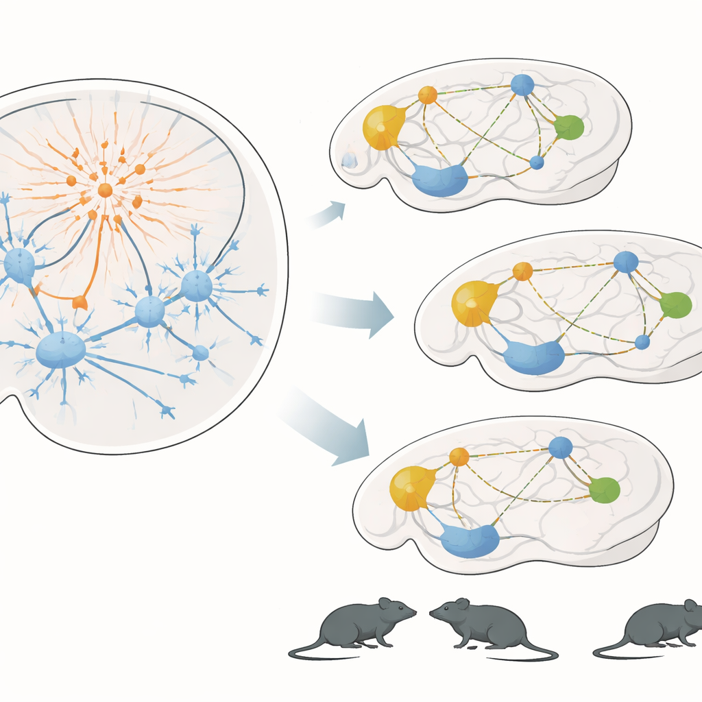
The team focused on the orbitofrontal cortex, a frontal brain region important for judging rewards and punishments, regulating emotions, and shaping social choices. Inside this region live parvalbumin interneurons—fast, inhibitory nerve cells that act as local timing and control units. Using a chemogenetic technique in male mice, the researchers engineered these cells to respond to a designer drug. Giving the drug briefly switched these interneurons into a highly active state, effectively turning down the output of neighboring excitatory cells in the orbitofrontal cortex without damaging the tissue.
Disrupted Brain Rhythms at Rest
To see how this local switch affected the whole brain, the scientists used high-resolution functional ultrasound imaging, which tracks changes in blood volume as a proxy for neural activity. They measured activity and coordination among 29 brain regions, with special attention to areas forming a rodent version of the human DMN, including hippocampus, thalamus, retrosplenial cortex, cingulate cortex, and the orbitofrontal cortex itself. After activating the orbitofrontal interneurons, connections between many of these regions weakened: key hubs such as the dorsal hippocampus, subiculum, thalamus, and retrosplenial cortex were less tightly linked in time. Some connections increased, hinting that when the main network is disturbed the brain partially reroutes traffic through alternative pathways, but the overall picture was one of reduced default-mode coordination.
From Brain Circuits to Social Life
The next question was whether this network disruption had visible effects on the animals’ lives. Groups of four mice lived together for days in a semi-natural arena equipped with automatic tracking. This setup allowed continuous monitoring of who approached whom, how often they sniffed each other, and how much they moved, without human interference. When the designer drug was given to mice with modified interneurons, they spent less time approaching and sniffing their cage-mates during the active night period, even though their overall movement stayed normal. In a separate memory test with familiar and new objects, these same mice explored as much as controls but failed to show a clear preference for the new object, suggesting a specific memory deficit rather than simple lethargy.
What This Means for Human Health
Taken together, the findings show that briefly over-activating a small group of inhibitory cells in the orbitofrontal cortex can ripple through the brain’s default network, weaken long-range communication, and produce both social withdrawal and memory problems in mice. This mirrors patterns seen in people with serious mental and neurodegenerative illnesses, where similar cell types and brain circuits are thought to be affected. While this study does not offer a treatment, it provides a mechanistic bridge between cell-level changes and complex social behavior. By pinpointing parvalbumin interneurons in the orbitofrontal cortex as important gatekeepers of social brain networks, the work suggests new directions for future research into how to protect or restore social functioning when these circuits go awry.
Citation: Khatamsaz, E., Ionescu, T.M., Keppler, K. et al. Orbitofrontal PV interneurons modulate social interaction via default mode network dynamics. Commun Biol 9, 573 (2026). https://doi.org/10.1038/s42003-026-10060-y
Keywords: social behavior, default mode network, orbitofrontal cortex, parvalbumin interneurons, functional connectivity