Clear Sky Science · en
Efficient cardiac MRI multi-structure segmentation for cardiovascular assessment with limited annotation by integrating data-level and network-level consistency
Why this research matters for heart health
Heart MRI scans can reveal life-saving details about how well the heart pumps, but turning these detailed images into numbers doctors can use usually requires time-consuming manual tracing by experts. This study introduces an artificial intelligence (AI) method that can learn to outline key parts of the heart accurately even when only a small fraction of the scans have been carefully labeled by specialists. By efficiently using the many unlabeled scans already stored in hospitals, the approach could make advanced heart analysis faster, cheaper, and more widely available.
Turning raw heart scans into useful measurements
To understand how a heart is working, cardiologists look at several structures in MRI images, including the main pumping chambers and the muscle wall. From these they calculate volumes, muscle mass, and how strongly the heart contracts—numbers that guide diagnosis, treatment choices, and long-term monitoring for conditions such as heart failure and cardiomyopathy. Today, these structures are often traced by hand or with software that still needs close supervision. The new method aims to largely automate this step, so that the outlines of the left ventricle, right ventricle, and surrounding heart muscle can be generated quickly and consistently for every patient.
Teaching computers with few examples
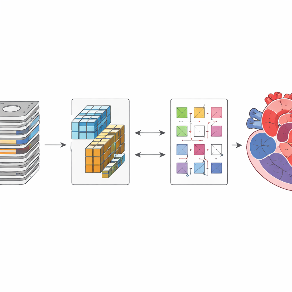
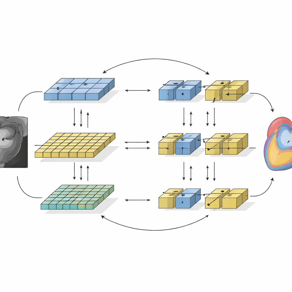
A major obstacle in building reliable medical AI is that the computer usually needs thousands of expertly labeled examples, and each heart scan can take hours to annotate. However, hospitals already hold large repositories of unlabeled images. The researchers tackled this by using a semi-supervised learning strategy, where a small set of labeled scans is combined with many unlabeled ones. The AI first learns from the labeled images in the usual way. At the same time, it is gently pushed to produce stable, self-consistent predictions on the unlabeled scans, so that even images without expert drawings still help refine the model. This approach reduces the reliance on costly manual work while still guiding the system toward anatomically sensible results.
Two different AI “viewpoints” working together
The framework, called SemiCoTr, marries two complementary types of image-analysis networks. One, based on traditional convolutional neural networks, is good at picking up fine local details. The other, built on more recent transformer technology, excels at seeing long-range relationships across the whole image. In SemiCoTr, both networks look at the same images and exchange information. Each network maintains a slowly updated “teacher” version whose predictions act as a stable reference for the rapidly changing “student” version, enforcing consistency over time. In addition, the two networks supervise one another: when one is confident about a structure on an unlabeled scan, its prediction helps train the other. This mutual guidance at both the data level and network level leads to more robust and anatomically coherent heart outlines.
How well does it actually work?
The team tested their method on a public heart MRI dataset of 100 patients used widely in the research community. They simulated a realistic scenario in which only 5–10% of the scans were labeled by experts and treated the rest as unlabeled. As more unlabeled data were added, the accuracy of the automatic segmentations rose steadily, approaching the performance of models trained on fully labeled datasets. Compared with several leading semi-supervised techniques, their approach consistently achieved higher accuracy and cleaner boundaries for all three key heart structures, while remaining computationally practical to train. Careful experiments showed that the gains came from both the multi-part loss design and the combination of the two different network types.
What this means for patients and clinics
The study shows that high-quality heart measurements from MRI can be obtained with far less manual annotation than was previously thought necessary. By making better use of the unlabeled scans that hospitals already generate every day, SemiCoTr points toward scalable AI tools that can deliver precise, reproducible heart assessments in busy clinical settings. In the long run, such systems could help doctors detect subtle heart problems earlier, tailor treatments more closely to individual patients, and track disease progression more reliably—all without adding to the workload of already stretched imaging specialists.
Citation: Guo, S., Zhao, X., Ren, J. et al. Efficient cardiac MRI multi-structure segmentation for cardiovascular assessment with limited annotation by integrating data-level and network-level consistency. npj Digit. Med. 9, 328 (2026). https://doi.org/10.1038/s41746-026-02475-y
Keywords: cardiac MRI, medical image segmentation, semi-supervised learning, deep learning, cardiovascular imaging