Clear Sky Science · en
Efficient SqueezeViT: A lightweight vision transformer framework for chest X-ray image classification
Why faster chest X-ray reading matters
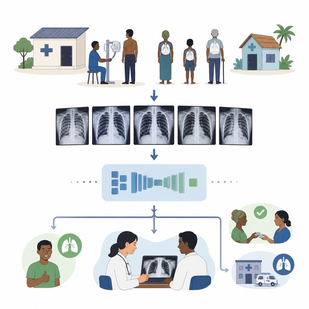
Chest X-ray scans are one of the most common ways doctors look for lung and heart problems, from pneumonia to tuberculosis. In busy hospitals or small clinics with limited computers, it is hard to run large artificial intelligence tools that could help doctors read these images quickly. This study presents a new compact AI model, called SqueezeViT, designed to spot chest diseases on X-rays while using far less computing power than typical systems, making it more practical for real-world care.
A new way to shrink smart image readers
Modern image-recognition tools often rely on two ideas. Convolutional neural networks are good at picking up fine details in small regions of an image, while transformer models are better at seeing the big picture across the entire scan. Standard vision transformers, however, are heavy and slow. The authors design SqueezeViT to keep the wide view of transformers but “squeeze” the amount of information that needs to be processed at each step. Their goal is to keep the parts of the image that matter for diagnosis while trimming away extra computation so the model can run on modest hardware.
How the compact model sees lungs and heart
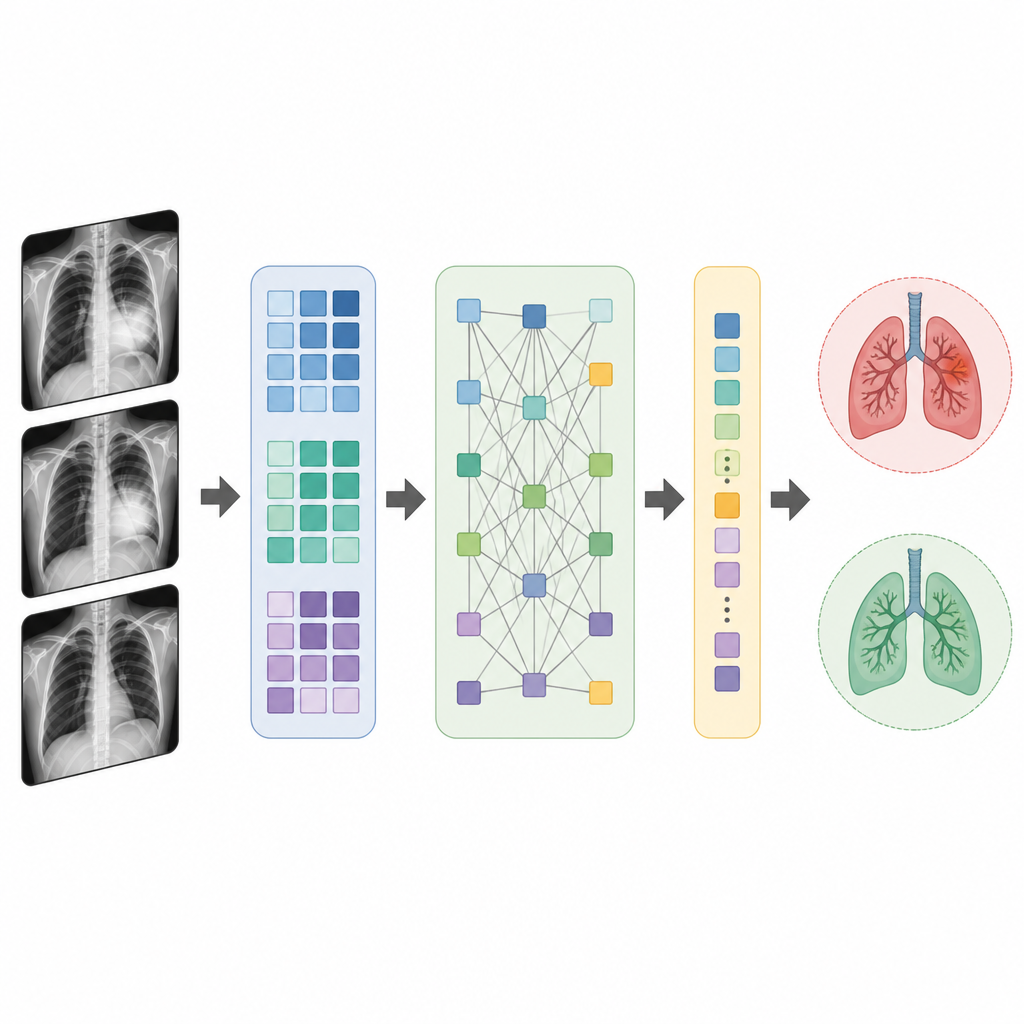
SqueezeViT combines two building blocks to handle chest X-rays efficiently. The first, called a Fire block, acts like a smart filter that compresses the information coming from the image into a smaller set of features, then expands it again to highlight patterns such as edges and textures tied to disease. The second, called the Translution Block, breaks the image into small patches and applies attention, allowing the model to relate signals from distant parts of the lungs or heart. By using slightly larger patches than many earlier designs, the model cuts the amount of work the attention step must do, while still capturing how changes in one part of the chest connect to others.
Putting the system to the test
To see how well SqueezeViT works in practice, the researchers evaluate it on two large public collections of chest X-rays: the NIH ChestX-ray14 dataset and the CheXpert dataset. Together, these include hundreds of thousands of images labeled for a range of conditions, such as cardiomegaly, edema, pneumonia, and lung nodules. The team trains SqueezeViT from scratch and compares its ability to distinguish sick from healthy cases against well-known deep learning models, including heavyweights like ResNet and DenseNet as well as lighter options like MobileNet, ShuffleNet, SqueezeNet, and MobileViT. They focus on the area under the receiver operating characteristic curve, a score that rewards models for ranking abnormal cases ahead of normal ones across different decision thresholds.
Speed, size, and accuracy in balance
The results show that SqueezeViT reaches accuracy on par with, and in several tasks better than, much larger models while being significantly smaller. It uses about half a million trainable parameters, cutting the parameter count by more than 40 percent compared with MobileViT and by over 90 percent compared with some of the largest baseline models. Its computations, memory use, and processing delays on both graphics processors and standard CPUs are all reduced, allowing it to analyze images in just a few milliseconds on typical hardware. In multi-disease settings, SqueezeViT matches or closely trails the best heavy models for many conditions and clearly surpasses other lightweight designs. For simple normal-versus-abnormal decisions, it again delivers strong and consistent scores across both datasets.
What this means for everyday care
For readers without a technical background, the key message is that SqueezeViT shows it is possible to build an AI assistant for chest X-rays that is both frugal with computing resources and careful in its disease detection. While it does not remove the need for radiologists or clinicians, it could help flag suspicious scans faster in crowded hospitals and extend advanced image analysis to clinics with limited equipment. The authors note that real-world labels can be noisy and some disease categories remain challenging, but they suggest that this compact design is a promising step toward reliable, portable support tools for chest imaging and may be adapted in the future to other scans such as CT or MRI.
Citation: Maurya, A., Lohia, A., Chirag et al. Efficient SqueezeViT: A lightweight vision transformer framework for chest X-ray image classification. Sci Rep 16, 16183 (2026). https://doi.org/10.1038/s41598-026-47918-4
Keywords: chest X-ray AI, vision transformer, medical image analysis, lightweight deep learning, lung disease detection