Clear Sky Science · en
Gene expression and machine learning techniques uncover corneal biomarkers associated with oxidative stress in the myopia progression
Why this matters for everyday eyesight
Myopia, or nearsightedness, is rising rapidly around the world, especially among young people who spend long hours on close-up tasks and screens. Most research has focused on the back of the eye, but this study turns the spotlight to the clear front window of the eye—the cornea. By probing tiny changes in corneal genes and immune activity linked to oxidative stress, the authors ask whether this tissue may quietly help drive myopia and whether it could one day guide new tests or treatments.
The eye’s front window under pressure
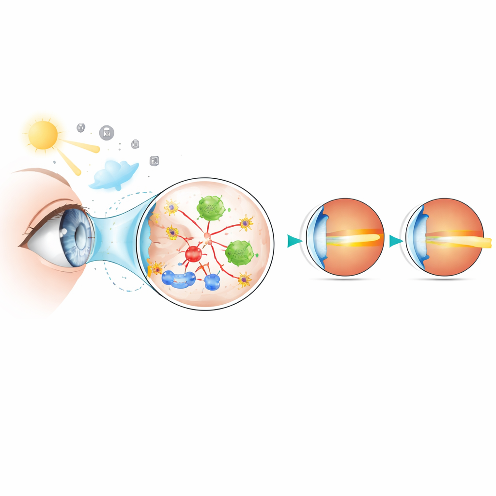
The cornea provides about two-thirds of the eye’s focusing power, so even subtle changes in its clarity or shape can alter vision. The researchers were interested in oxidative stress, a biochemical imbalance in which harmful oxygen byproducts overwhelm the body’s defenses. Earlier studies suggested that oxidative stress is present in eye fluids and the retina of myopic eyes, but the cornea had received little attention. Because it is easily accessible during routine eye surgery and treatments, the team reasoned that the cornea could be both a sensor of damaging stress and a practical source of biomarkers—measurable molecular signs of disease.
Mining big data to find molecular fingerprints
To search for such fingerprints, the scientists combined several publicly available gene expression datasets from corneal tissue and lens cells of people with and without myopia. They first identified thousands of genes whose activity differed between myopic and non-myopic samples, then narrowed this list to a smaller group already known to be related to oxidative stress. Using modern machine learning techniques, including LASSO regression and the Boruta feature-selection algorithm, they homed in on three key genes—ATF3, GRIN2B, and GSTM3—that consistently distinguished myopic tissue from normal tissue.
Three warning lights in the cornea
ATF3 helps cells respond to stress signals, GRIN2B is part of a communication channel usually studied in the brain, and GSTM3 helps detoxify reactive molecules using the antioxidant glutathione. In both combined datasets and an independent validation set, all three markers were expressed at lower levels in myopic samples. The team then checked real human corneal tissue removed during SMILE laser surgery from young adults with low and high myopia. They found that ATF3 and GSTM3 messenger RNA levels were significantly lower in highly myopic corneas, and protein measurements confirmed that all three markers were reduced. Intriguingly, a statistical technique called Mendelian randomization did not support these genes as direct genetic causes of myopia, hinting instead that myopia or its environment may suppress their activity.
Immune shifts and stressed cell pathways
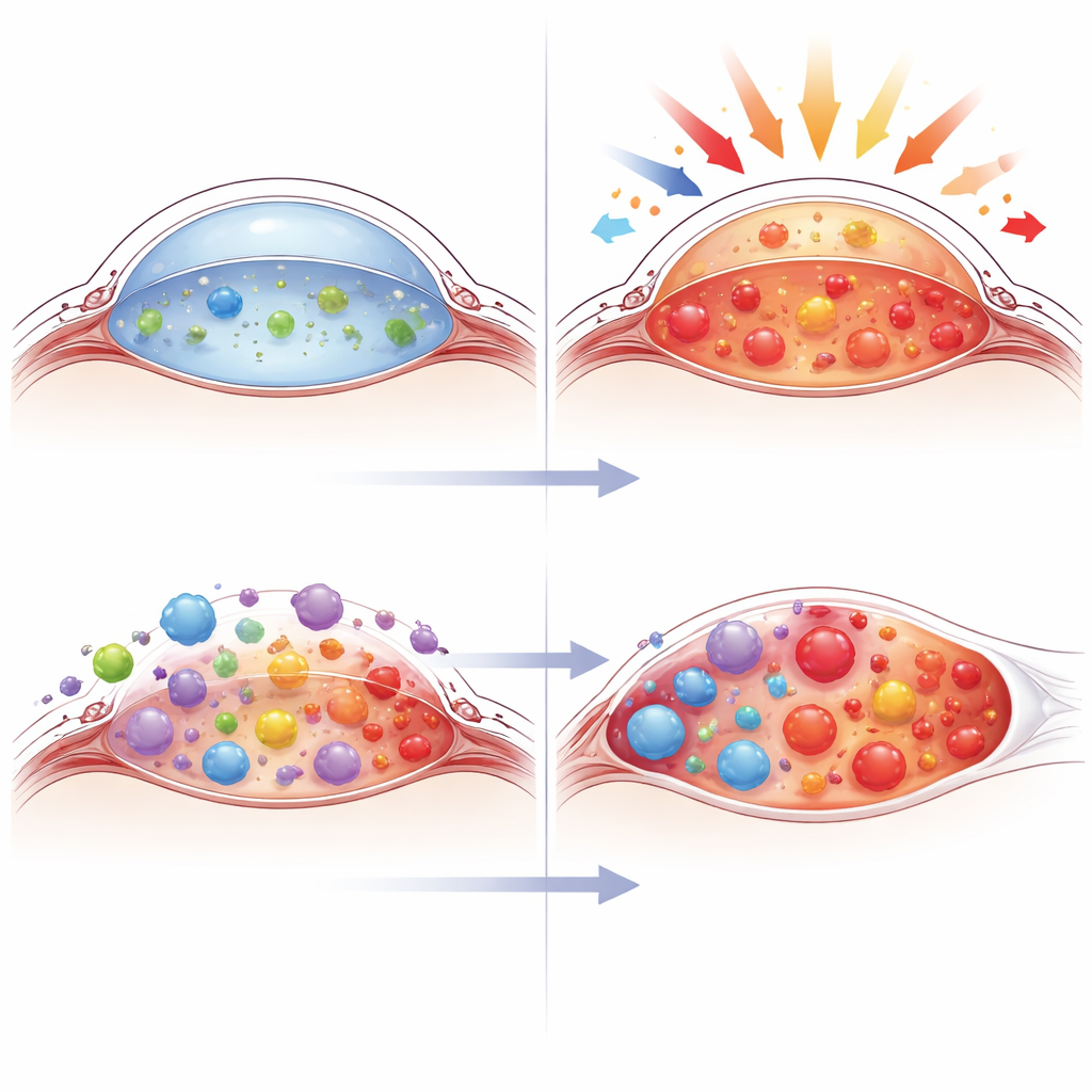
Looking beyond single genes, the authors examined how immune cells and broader cellular pathways differed between groups. They estimated the mix of 22 immune cell types present in corneal samples and found notable changes in individuals with myopia, including more CD8+ T cells and monocytes and fewer eosinophils and resting memory CD4+ T cells. The three biomarkers were strongly negatively linked to CD8+ T cells and positively linked to eosinophils, suggesting that reduced oxidative stress defenses go hand in hand with a more inflammatory immune landscape. Additional pathway analyses tied the biomarkers to cell-to-cell junctions, responses to low oxygen, and damage from ultraviolet light. Together, these findings support a picture in which chronic stress in the corneal microenvironment reshapes both cell signaling and immune activity.
Hints of future treatments and current limits
To explore therapeutic angles, the researchers used drug–gene databases and computer-based molecular docking to predict compounds that might interact with the three biomarkers. Retinoic acid, an active form of vitamin A important for corneal health, emerged as a shared candidate that could, in theory, influence all three. However, these predictions are purely computational and require careful laboratory and clinical testing. The authors also stress that their study relies on relatively small sample sizes and mixed tissue sources, and that many of the inferred mechanisms remain to be proven experimentally.
What this means for people with myopia
Overall, the study suggests that the front of the eye is not just a passive lens but an active participant in myopia, with oxidative stress–related genes and immune cells shifting in ways that track with disease severity. ATF3, GRIN2B, and GSTM3 stand out as promising biomarkers that may one day help doctors assess myopia risk or monitor progression using easily obtained corneal tissue. While it is too early to change clinical practice, this work lays a molecular foundation for future strategies aimed at protecting the cornea’s antioxidant defenses and calming harmful immune responses to slow or prevent worsening nearsightedness.
Citation: Zhou, Q., Ye, M., Zhang, Z. et al. Gene expression and machine learning techniques uncover corneal biomarkers associated with oxidative stress in the myopia progression. Sci Rep 16, 10651 (2026). https://doi.org/10.1038/s41598-026-46896-x
Keywords: myopia, cornea, oxidative stress, biomarkers, ocular immunity