Clear Sky Science · en
Ubiquitination-driven fibroblast dysfunction: a multi-omics blueprint for precision diagnosis and therapy in diabetic foot ulcer
Why stubborn foot wounds matter
For many people living with diabetes, a small sore on the foot can turn into a stubborn ulcer that refuses to heal. These diabetic foot ulcers often lead to long hospital stays and even amputation, yet doctors still struggle to predict which wounds will worsen and which treatments will work best. This study digs deep into the cells inside these chronic wounds and uncovers a hidden control system that may explain why healing breaks down—and how we might diagnose and treat these ulcers more precisely in the future. 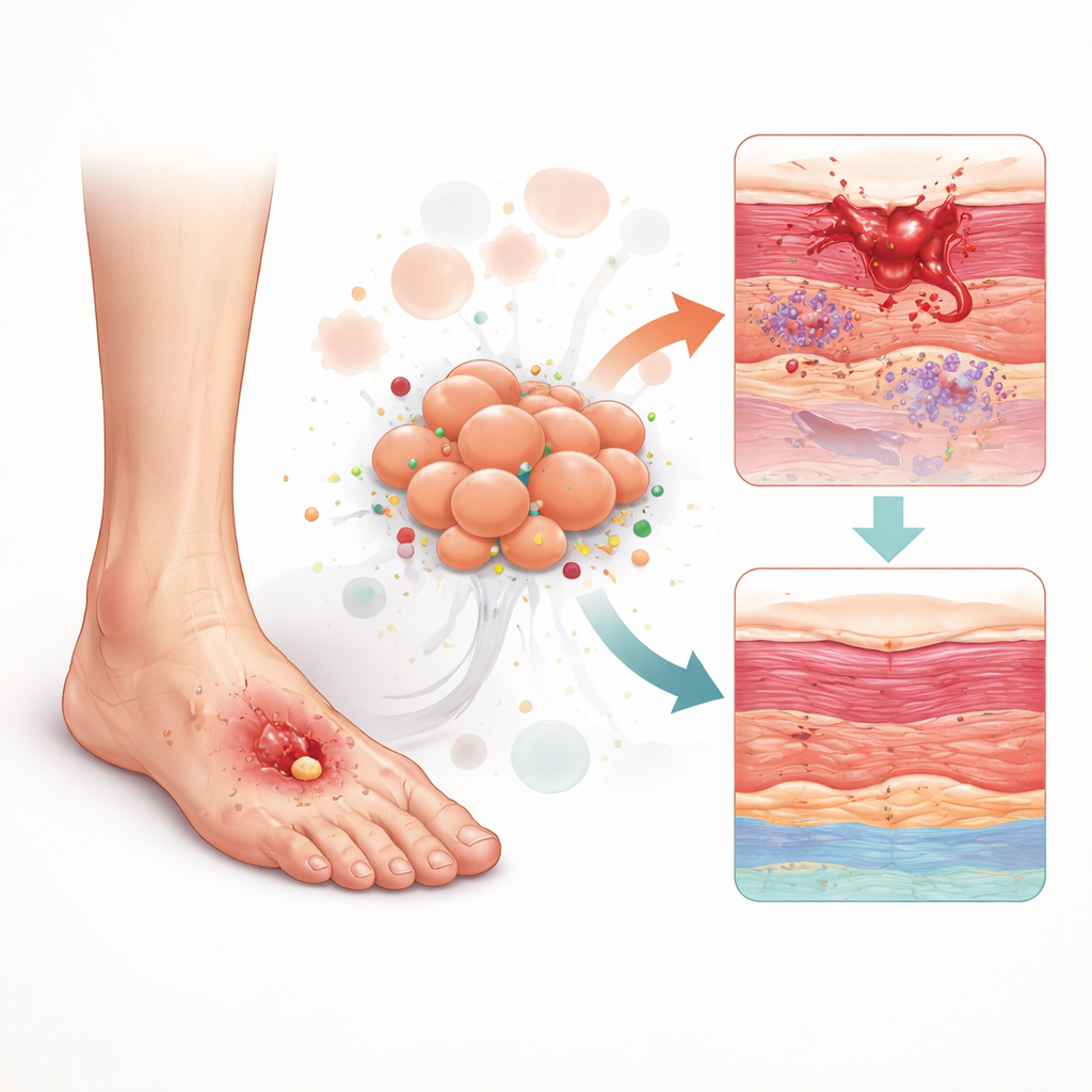
A closer look at the skin’s repair crew
Healthy skin relies on a team of cells that patch up damage after an injury. Among the most important are fibroblasts, the structural “handymen” that build and remodel the tissue scaffolding during wound repair. The researchers used cutting-edge single-cell RNA sequencing to analyze more than 23,000 individual cells from normal foot skin and diabetic foot ulcers. This allowed them to see which genes were switched on in each cell type, from immune cells and blood vessel cells to fibroblasts. They focused on a group of genes involved in ubiquitination—a chemical tagging system that marks proteins for recycling or relocation and helps keep cellular machinery in balance.
Discovery of a troublemaking fibroblast group
When the team compared healthy and diseased tissues, fibroblasts emerged as the most disrupted cell type in diabetic ulcers. A distinct subgroup of fibroblasts appeared almost exclusively in ulcer tissue. These “pathogenic” fibroblasts showed signs of higher stem-like potential, altered metabolism, and rewired communication with neighboring cells. Using computational tools that trace how cells change over time, the study suggested that ordinary fibroblasts gradually shift into this harmful state in the harsh diabetic wound environment. Once transformed, they send and receive excessive signals related to inflammation, new blood vessel growth, and tissue remodeling—signals that, instead of promoting repair, may lock the wound into a chronic, inflamed state. 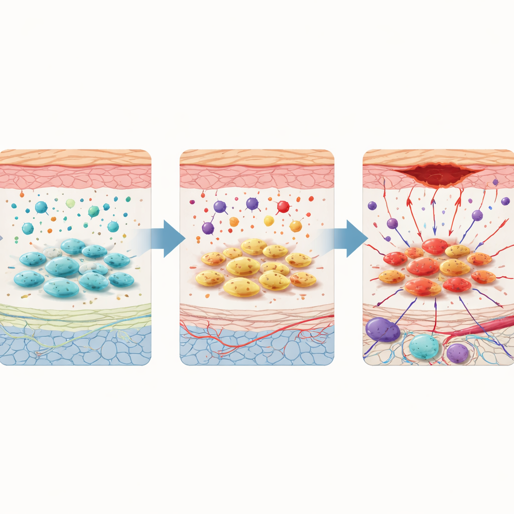
Hidden molecular switches and a diagnostic fingerprint
To turn thousands of gene measurements into something clinically useful, the researchers combined several machine learning methods to sift through large public datasets of diabetic and healthy foot tissues. They identified four key genes—MEF2A, SKIL, MAF, and KRT5—that together formed a powerful “fingerprint” distinguishing diabetic ulcers from normal skin, with strong accuracy in test data. Among these, SKIL stood out as the most influential. It is known from other research to interfere with a major repair pathway (often controlled by TGF-β) that guides fibroblasts in building new tissue. In the diabetic ulcers, SKIL was consistently overactive and tightly linked to shifts in how fibroblasts use energy, especially increased reliance on sugar-burning pathways such as glycolysis.
Inflamed defenses and tailored disease subtypes
The study also examined the immune cells that infiltrate diabetic foot ulcers. Compared with normal skin, diabetic ulcers contained more inflammatory cells—such as certain macrophages and neutrophils—and fewer regulatory cells that normally help cool down the response. By grouping ulcer samples based on the activity of ubiquitination-related genes, the researchers uncovered two distinct molecular subtypes: one dominated by inflammation and metabolism-related changes, and another enriched for signals involved in blood vessel growth and tissue scarring. These patterns suggest that not all ulcers are the same at the molecular level, which may explain why patients respond differently to the same treatments and highlight the need for personalized care.
New drug possibilities hiding in plain sight
Because SKIL appeared central to fibroblast dysfunction, the researchers searched drug–gene databases and used molecular docking simulations to predict medicines that might bind to and influence SKIL. They identified two candidates: lumicolchicine and ramipril. Ramipril is especially intriguing because it is an existing blood pressure medication commonly prescribed to people with diabetes. The simulations suggest that ramipril could interact with SKIL while also improving blood vessel function, hinting that this familiar drug class might one day be repurposed to help diabetic wounds heal more effectively. These predictions still require laboratory and clinical testing, but they open a practical path toward translating molecular insights into therapy.
What this means for patients and care
Taken together, this work maps how a protein-tagging system inside fibroblasts can drive them off course, fueling chronic inflammation and poor repair in diabetic foot ulcers. By pinpointing a specific harmful fibroblast subtype, defining a four-gene diagnostic signature, and highlighting SKIL as a potential drug target, the study offers a blueprint for more precise diagnosis and individualized treatment. In the long run, such molecularly informed strategies could help clinicians identify high-risk ulcers earlier, tailor therapies to the biology of each wound, and perhaps use existing drugs in new ways to prevent limb-threatening complications.
Citation: Wang, W., Peng, X., Hua, Q. et al. Ubiquitination-driven fibroblast dysfunction: a multi-omics blueprint for precision diagnosis and therapy in diabetic foot ulcer. Sci Rep 16, 14669 (2026). https://doi.org/10.1038/s41598-026-45436-x
Keywords: diabetic foot ulcer, fibroblasts, ubiquitination, wound healing, precision medicine