Clear Sky Science · en
Establishment of a xenograft model of endometriosis-associated fibrosis using human immortalized endometrial stromal cells overexpressing HOXC8
Why this research matters for women’s health
Endometriosis affects millions of women worldwide, often causing severe menstrual pain, chronic pelvic discomfort, and infertility. A major but less visible part of the disease is scarring and internal “sticking together” of tissues, known as fibrosis and adhesions, which can distort organs and make symptoms hard to treat. Yet doctors currently have no therapies that directly target this scarring. This study describes a new laboratory model that mimics the fibrotic, scar-forming side of endometriosis, opening the door to testing medicines that might finally prevent or reverse this damage.
From monthly pain to lasting scars
Endometriosis occurs when tissue similar to the uterine lining grows outside the uterus, where it does not belong. These misplaced patches of tissue bleed and inflame the surrounding area, and over time the body responds by laying down collagen fibers, the same material found in scars. This gradual build-up turns flexible tissue into stiff bands and sheets that can tether organs together. Current treatments mainly suppress hormones or ease pain, but they do not specifically stop scar formation, and they are often unsuitable for women trying to become pregnant. One reason progress has been slow is the lack of animal models that faithfully reproduce the fibrotic aspect of endometriosis rather than just the presence of misplaced tissue.
A suspect gene comes into focus
The researchers previously used large-scale gene analyses and computer modeling to search for “master switches” that might drive many of the gene changes seen in endometriosis. One candidate was a developmental gene called HOXC8, which is abnormally active in endometriotic tissue and is also implicated in several cancers and in liver scarring. Earlier work in primary cell cultures showed that turning up HOXC8 boosted the cells’ ability to move, invade, and contract collagen gels, suggesting a pro-scarring role through a well-known signal relay called the TGFB/SMAD pathway. But these findings were all in dishes; the key question was whether HOXC8 could actually promote fibrosis inside a living body in a way that resembles endometriosis.
Building a scar-forming model in mice
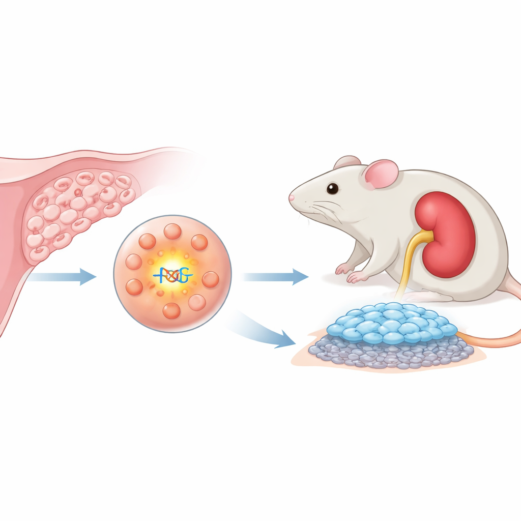
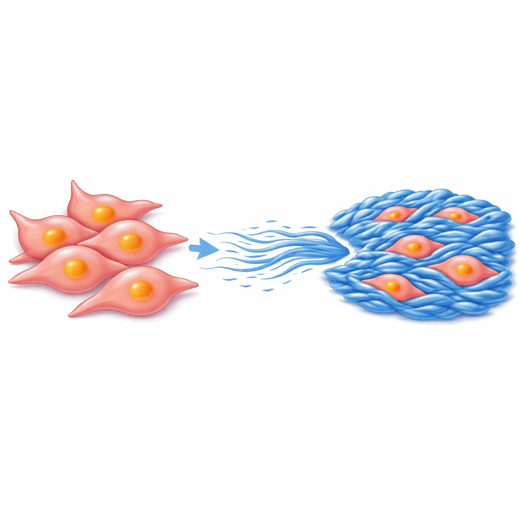
To test this, the team engineered immortalized human endometrial stromal cells—the type of cells that form the supporting framework of the uterine lining—to produce extra HOXC8. They also created matched control cells without this extra gene activity. In laboratory tests, the HOXC8-high cells did not divide faster, but they did move, invade through a gel, close artificial “wounds,” and contract collagen more strongly than control cells, all behaviors associated with aggressive, scar-forming tissue. The scientists then mixed these cells into a collagen gel to form small aggregates and transplanted them under the thin outer capsule of the kidneys of highly immunodeficient mice, a protected niche that allows human cells to survive and form lesions.
How a single switch drives scarring
Five weeks after transplantation, both types of human cells had formed visible lesions on the mouse kidneys, confirming they could engraft and persist. However, lesions derived from HOXC8-overexpressing cells were thicker and richer in collagen fibers, as shown by special blue staining and by strong signals for a major collagen protein, COL1A1. Both control and HOXC8 lesions contained myofibroblast-like cells—the usual culprits in fibrosis—but only the HOXC8 lesions accumulated large amounts of collagen, suggesting that HOXC8 changes how these cells behave rather than just how many appear. Importantly, within the HOXC8 lesions, the TGFB/SMAD signaling proteins SMAD2 and SMAD3 were found in their activated, phosphorylated form inside cell nuclei, both in culture and in the mouse grafts, linking HOXC8 activity directly to a known fibrosis-driving pathway in living tissue.
Toward targeted anti-scarring therapies
The study’s main contribution is the creation of a reproducible mouse xenograft model in which human endometrial stromal cells, pushed into a HOXC8 “on” state, reliably form collagen-rich, endometriosis-like fibrotic lesions. Because the model depends on a defined molecular switch and a specific signaling route, it provides a controlled platform to probe how fibrosis arises and to test drugs that block key steps, such as inhibitors of the TGFB receptor ALK5 that have already shown promise in cell culture. For patients, this does not yet translate into a new treatment, but it represents a crucial step: researchers now have a realistic, human-cell–based system for studying the scar-forming heart of endometriosis and for exploring therapies aimed at preserving pelvic organs before irreversible damage occurs.
Citation: Takasaki-Kawasaki, H., Sato, S., Tamehisa, T. et al. Establishment of a xenograft model of endometriosis-associated fibrosis using human immortalized endometrial stromal cells overexpressing HOXC8. Sci Rep 16, 11318 (2026). https://doi.org/10.1038/s41598-026-41956-8
Keywords: endometriosis, fibrosis, HOXC8, TGFB SMAD signaling, xenograft model