Clear Sky Science · en
Research on characteristic recognition and quantification of internal powder residue in LPBF porous structure based on image processing
Why hidden powder in 3D-printed parts matters
Metal 3D printing is rapidly moving from the lab into airplanes, cars, and even bone implants. But inside many of these intricate, sponge-like parts, leftover powder grains can remain trapped and unseen, weakening the structure and risking failure in service. This paper presents a new way to "see" and measure that hidden powder automatically, using advanced X-ray imaging and smart image processing, with the goal of making 3D-printed metal parts safer and more reliable.
The promise and problem of porous metal parts
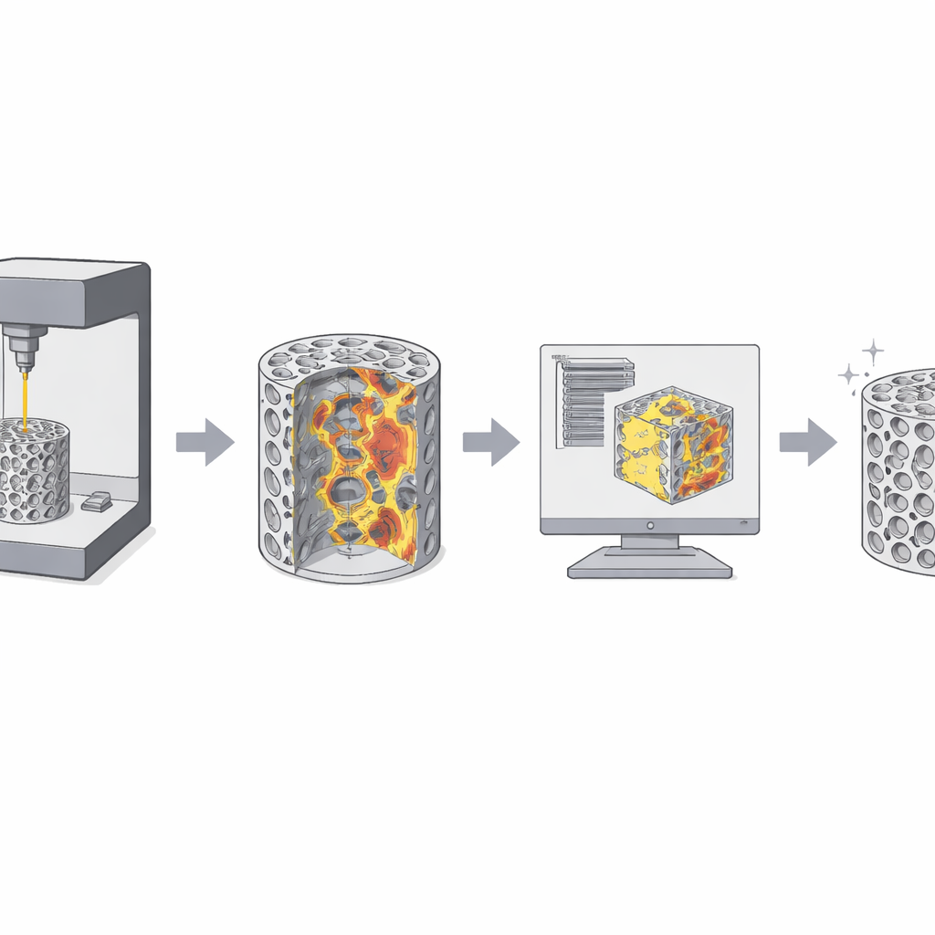
Laser Powder Bed Fusion (LPBF) builds metal components layer by layer from fine powder, enabling complex porous shapes that are ideal for lightweight structures and bone-mimicking implants. In this study, the authors focus on porous zinc scaffolds whose outer ring imitates dense bone and whose inner region imitates the spongy interior. However, in such maze-like interiors the laser cannot fully reach every nook and cranny, and tiny powder particles can remain unmelted or only partially fused to the walls. These residues can clog fluid channels, concentrate stress, disrupt how the material slowly dissolves in the body, and even provoke inflammation. Detecting and quantifying this trapped powder deep inside the solid metal is therefore essential for both mechanical safety and biological performance.
Looking inside with X-rays and digital slices
Among non-destructive inspection methods, X-ray computed tomography (CT) stands out because it can see inside dense metals and reconstruct a full three-dimensional view. The researchers scanned cylindrical zinc scaffolds at micrometer resolution, slicing the 3D volume into thousands of 2D images. They chose cross-sections along the direction that best revealed the repeating internal pattern. On these slices, metal, empty pores, and residual powder appear as regions with different gray levels. But manually going through over a thousand images, setting thresholds by eye in commercial software, is slow, subjective, and often inaccurate—especially when image contrast is low or brightness drifts from slice to slice.
Teaching a computer to find the powder
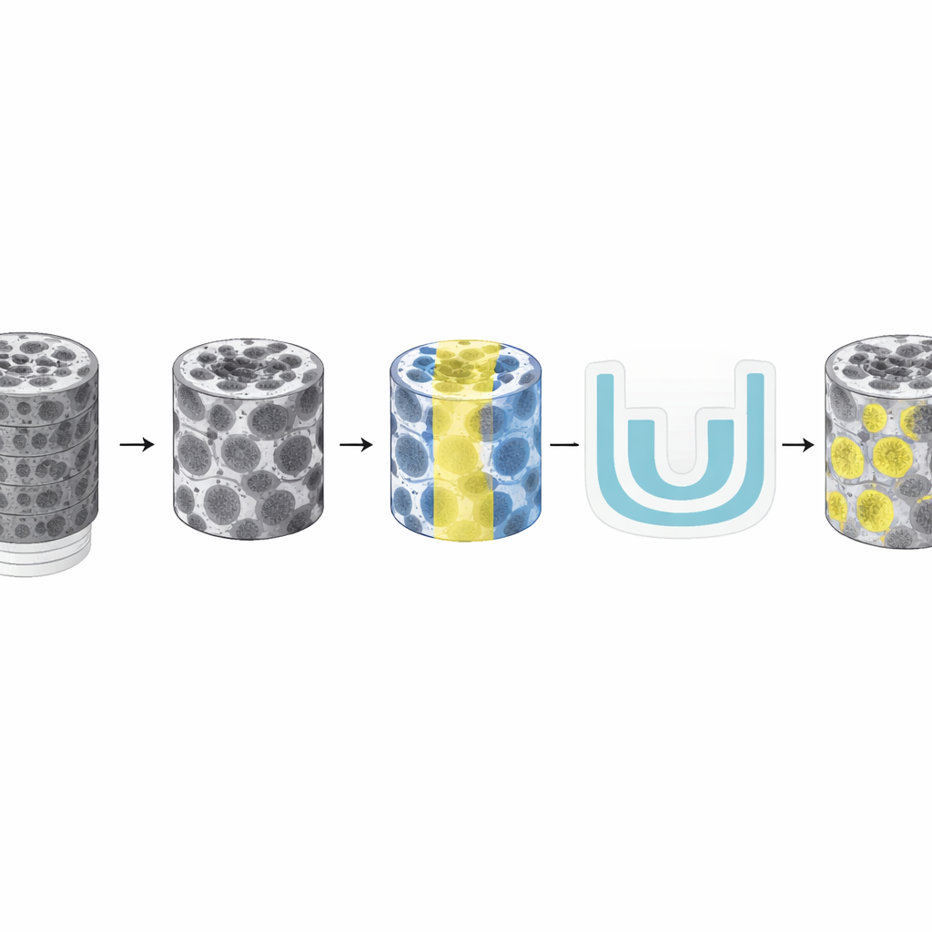
To overcome these limitations, the team built an automated detection pipeline in MATLAB that combines classical image processing with deep learning. First, each CT slice is carefully preprocessed: the images are standardized to a common format, cropped to remove empty background, denoised, contrast-enhanced, and sharpened so that pores, solid skeleton, and powder grains stand out more clearly. Then a newly designed dual-threshold Otsu algorithm scans the gray-level histogram and automatically splits each pixel into three categories: open pore, solid framework, or candidate powder. By using two thresholds instead of one, the method better separates subtle gray differences between solid metal and stuck powder, which often overlap in brightness.
Adding deep learning for fine detail
Thresholding alone still struggles with fuzzy edges and low-contrast regions, so the authors train a U-Net, a deep learning model widely used for medical imaging. They first generate draft labels with their dual-threshold method and then have an expert refine around 300 representative slices by hand, creating high-quality examples of what is and is not residual powder. The U-Net learns from these examples to recognize powder patterns in the CT slices, including free particles, semi-molten grains partly attached to the walls, and larger bonded clusters. In the final system, the dual-threshold step provides a coarse map, and the U-Net cleans up the boundaries and corrects subtle mistakes. Morphological operations further remove tiny noise spots while preserving meaningful agglomerates.
From images to decisions on cleaning
Once the powder regions are segmented, the software reconstructs their 3D shapes and measures properties such as volume, location, and particle size distribution, while also computing overall porosity. Compared with a pycnometer fluid-displacement measurement, the CT-based porosity values agree closely, and the new method clearly outperforms both the popular ImageJ workflow and fully manual inspection. Processing 1,463 CT images takes about 12 minutes with the MATLAB pipeline, versus 4 hours in ImageJ and 6 hours by hand, while recognition accuracy reaches roughly 86–89%. The authors also stress-tested the system by varying thresholds and adding noise, and found that the combined dual-threshold-plus-U-Net approach remains robust. Finally, they link the 3D powder maps to simple rules that recommend suitable cleaning strategies, such as ultrasonic vibration for loose powder deep in channels or chemical treatment plus blasting for more strongly attached grains.
What this means for safer 3D-printed parts
In plain terms, the study shows how to turn stacks of gray X-ray slices into practical guidance for cleaning and qualifying complex 3D-printed metal parts. By automatically spotting where and how much powder remains inside porous structures, and by doing so far faster than a human expert, the method lays the groundwork for an integrated "detect-identify-clean" toolkit. While the current work focuses on regularly patterned zinc scaffolds, the underlying idea—using CT gray-level differences and machine learning to track residual powder—could be extended to other metals and shapes. As metal 3D printing moves into critical implants and high-performance components, such intelligent inspection pipelines will be key to ensuring that what we cannot see inside does not compromise safety and function.
Citation: Shi, W., Cao, S., Hou, Q. et al. Research on characteristic recognition and quantification of internal powder residue in LPBF porous structure based on image processing. Sci Rep 16, 13247 (2026). https://doi.org/10.1038/s41598-026-40479-6
Keywords: laser powder bed fusion, porous metal scaffolds, computed tomography, image segmentation, residual powder