Clear Sky Science · en
Safety and biodistribution of intrathecal administration of mesenchymal stem cells (MSCs) and neurotrophin-releasing nanoparticles in a porcine CSF-guided delivery model for amyotrophic lateral sclerosis (ALS) drug discovery
New Ways to Deliver Help for Failing Nerves
Amyotrophic lateral sclerosis (ALS) is a devastating disease that slowly paralyzes people by killing the nerve cells that control movement. Current drugs only modestly slow its progress. Scientists are searching for therapies that can better protect these vulnerable nerve cells. This study explores a two-part experimental treatment in pigs: healing cells known as mesenchymal stem cells and tiny particles that slowly release nerve-nourishing proteins. The work does not test whether the treatment cures ALS, but instead asks a simpler, crucial question: is this approach safe, and where do the cells go in the body after they are injected near the spinal cord?
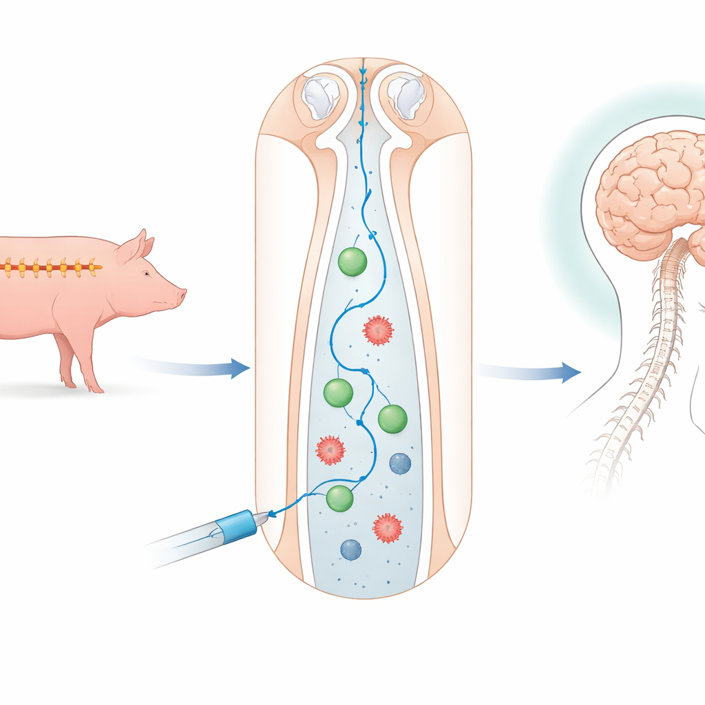
Why Pigs and Spinal Fluid Matter
Most ALS experiments start in mice, but their bodies are quite different from ours, especially in size and in the structure of the nervous system. Pigs, by contrast, have a spine and brain more similar in scale to humans, making them a valuable bridge between rodent studies and human trials. In this study, twelve young male pigs were divided into four groups. Some received only salt solution, some received stem cells from human fat tissue, others received stem cells from the jelly-like tissue of umbilical cords, and one group underwent spinal puncture without any injection. All injections were given into the fluid-filled space surrounding the spinal cord in the lower back, the same route doctors sometimes use to deliver medicines to human patients. Later, the pigs also received nanoparticles loaded with two nerve-supporting proteins.
Tiny Helpers: Stem Cells and Nerve-Nurturing Particles
The stem cells used here are not intended to turn into new nerve cells directly. Instead, they act more like mobile pharmacies, releasing substances that calm inflammation and support surviving nerve cells. The nanoparticles carry two natural proteins, brain-derived neurotrophic factor (BDNF) and neurotrophin‑3 (NT3), which help nerve cells survive and maintain their connections. To keep these fragile proteins from breaking down too quickly, the team wrapped them in a protective coat of a common, body-friendly polymer called PEG. Careful lab tests showed that these coated particles stayed stable in size, charge, and protein content for nearly a month, suggesting they can provide a steady, gentle release of the helpful proteins rather than a short-lived burst.
Tracking Safety Inside Blood and Brain
To judge safety, the researchers monitored the pigs for three weeks. They repeatedly tested blood and spinal fluid, paying particular attention to C‑reactive protein, a general marker of inflammation. Most blood counts and organ-related measures stayed within normal limits or showed only brief, scattered changes not clearly tied to treatment. One pig in the fat‑derived cell group died during recovery from anesthesia, but detailed examination did not link this to the cells or nanoparticles. Interestingly, levels of C‑reactive protein tended to drop after the nanoparticle dose, especially in the groups that received stem cells, hinting that this combined approach may soften inflammation rather than stir it up. Magnetic resonance imaging scans of the spine taken before, during, and after procedures showed no structural damage or worrisome changes in any group.
Where the Transplanted Cells Travel
Another key question was where the human stem cells move once they are released into the spinal fluid. To follow them, the scientists labeled the cells with tiny iron particles that show up as dark spots on MRI scans and can be detected by special tissue stains. Imaging during injection revealed clear signals in the spinal canal above the entry site, confirming that the cells were indeed delivered into the fluid space. Later tissue studies showed iron deposits around the outer surfaces of the spinal cord and brain, especially near fluid-filled cavities, but not deep inside the nerve tissue itself. This pattern suggests that the cells stay in the spaces bathed by spinal fluid and influence nearby nerve cells from the outside by releasing helpful substances, rather than burrowing into and replacing damaged cells.
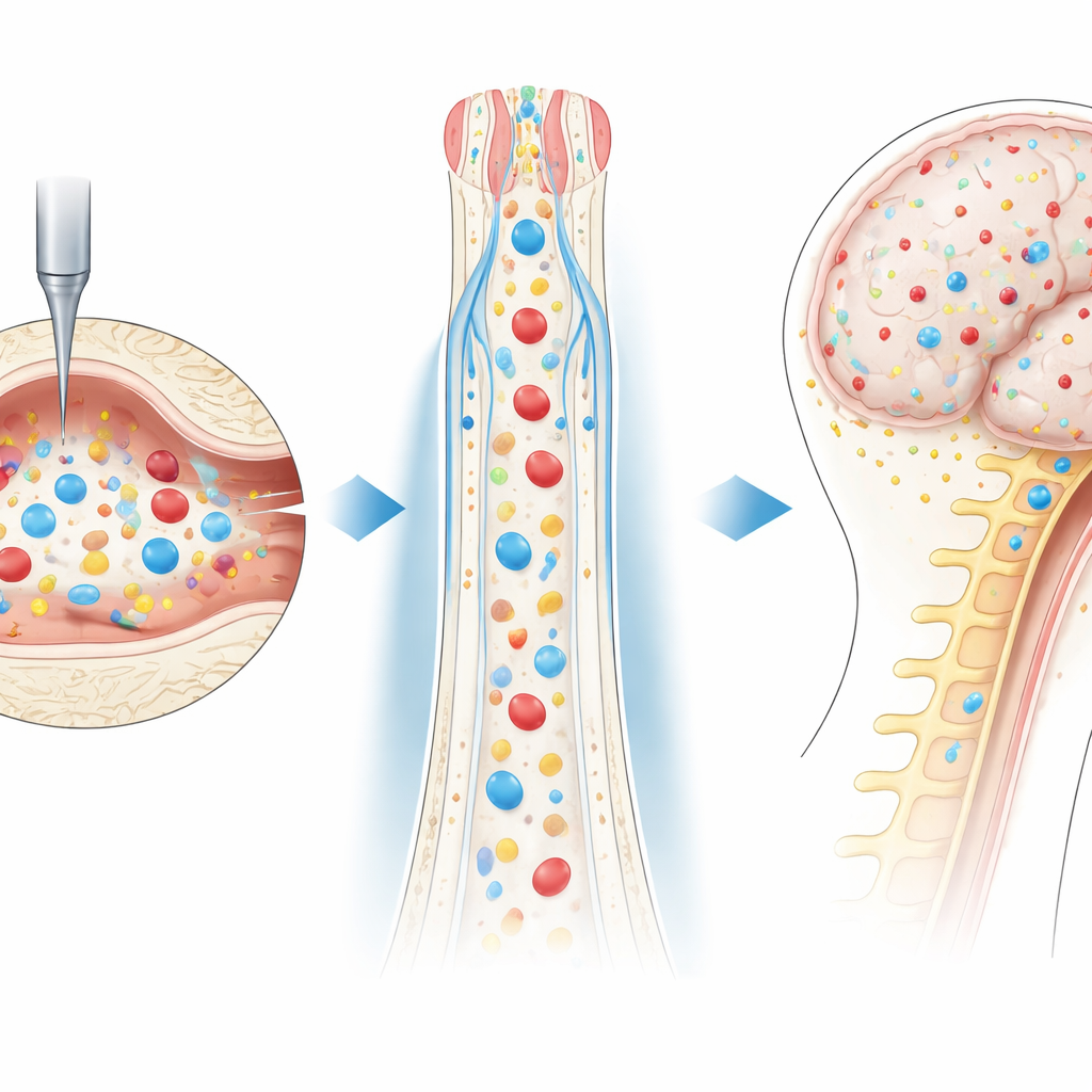
What This Means for Future ALS Treatments
This work does not prove that stem cells and neurotrophin‑releasing nanoparticles can halt ALS in patients. The pigs were healthy, and the study focused only on short‑term safety and on where the cells and particles travel. Still, the findings are encouraging: repeated injections into the spinal fluid were well tolerated, vital organs remained unharmed, inflammation markers trended downward after treatment, and the introduced cells stayed confined to the fluid spaces around the brain and spinal cord. Together, these results build an early but important foundation for future trials in animals that actually develop ALS‑like disease and, eventually, for carefully designed studies in people. The approach offers a way to bathe threatened nerve cells in a sustained, protective environment—potentially turning the body’s own repair systems into powerful allies against neurodegeneration.
Citation: Sinderewicz, E., Dąbkowska, M., Sarnowska, A. et al. Safety and biodistribution of intrathecal administration of mesenchymal stem cells (MSCs) and neurotrophin-releasing nanoparticles in a porcine CSF-guided delivery model for amyotrophic lateral sclerosis (ALS) drug discovery. Sci Rep 16, 11216 (2026). https://doi.org/10.1038/s41598-026-40196-0
Keywords: amyotrophic lateral sclerosis, mesenchymal stem cells, neurotrophins, nanoparticle drug delivery, intrathecal therapy