Clear Sky Science · en
Hybrid deep neural network with PCA based features optimization for enhancing brain tumor classification
Why spotting brain tumors early matters
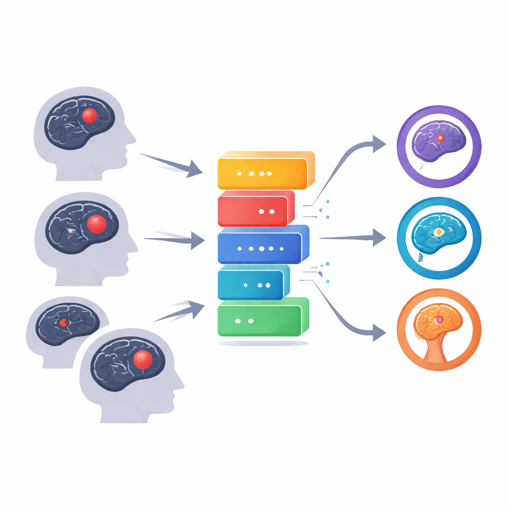
Brain tumors are among the most feared cancers because they grow inside the body’s command center, often silently, until they cause serious symptoms. Doctors rely on MRI scans to find and track these tumors, but modern scanners generate thousands of detailed images for each patient. No matter how skilled, a human radiologist can struggle to sift through all that information quickly and consistently. This study describes a computer system that helps by reading brain scans and sorting them into four groups—glioma, meningioma, pituitary tumor, or no tumor—with accuracy that rivals or exceeds many previous methods.
Turning complex scans into clearer pictures
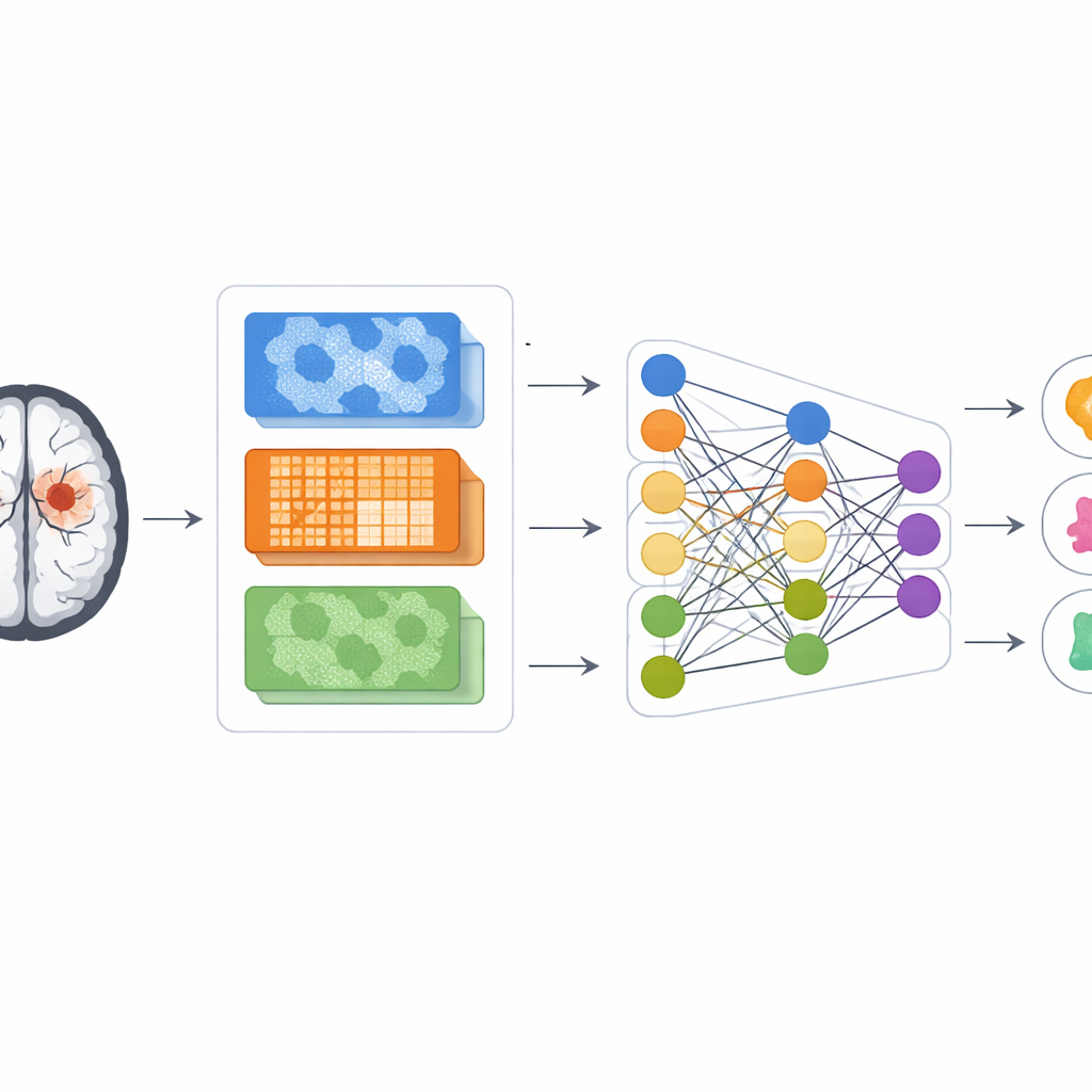
At the heart of the work is an effort to tame the flood of visual data coming from MRI machines. The authors build on a powerful image-recognition network called DenseNet121, originally trained on everyday photographs, and retrain it to recognize patterns in brain scans. Before the images ever reach this network, they are carefully prepared: their size is standardized, contrast is smoothed and enhanced, and the regions most likely to contain tumors are isolated using a clustering method that groups similar pixels together. These steps help the computer focus on the brain tissue that matters most and reduce distractions from noise or background structures.
Blending several ways of looking at a tumor
Rather than relying only on what the deep network learns by itself, the system also computes classic image descriptors that radiologists and engineers have used for years. One set captures how bright and dark spots are arranged next to each other, highlighting texture and edges. Another focuses on very small local patterns in pixel intensity, which can reveal subtle graininess in tissue. A third follows how similar intensities cluster together across larger areas, emphasizing whether a region is smooth and uniform or patchy and irregular. By fusing these three viewpoints into a single feature set, the model gets a richer description of each suspected tumor than from deep learning alone.
Making the data manageable and trustworthy
The combined description of each scan is extremely high‑dimensional, which can confuse a learning algorithm and lead to overfitting—where the model memorizes training images instead of learning general rules. To avoid this, the authors use a mathematical tool called principal component analysis to compress the information into a smaller number of informative components before feeding it into the classification network. Importantly, all of these steps are built to mimic real clinical use: scans from each patient are kept either in the training group or the testing group, never both, so the model cannot cheat by seeing the same anatomy twice. The network itself includes dropout layers and data shuffling, techniques that deliberately add randomness during training so that the final system is more robust to new cases.
How well the system recognizes different tumors
The researchers test their method on more than seven thousand MRI images from a widely used public collection. The scans cover four categories: glioma, meningioma, pituitary tumor, and normal brain. Using their hybrid design, they reach about 95.9% overall accuracy. Precision, recall, and the combined F1 score—all standard measures of how many cases are correctly labeled and how many errors are made—each stay around 94%. The model is particularly strong at identifying pituitary tumors and normal brains, and only rarely confuses gliomas with meningiomas, which can look similar at their borders. Learning curves show that performance on training and unseen validation images rises together without large gaps, suggesting that the network has avoided the common pitfall of overfitting.
What this could mean for patients and doctors
For a non‑specialist, the main message is that combining several ways of describing an image with a modern deep learning network can turn raw MRI scans into reliable, automated suggestions about tumor type. The system is not meant to replace radiologists, but to serve as a second pair of eyes that works quickly and consistently across large numbers of scans. With further testing on data from different hospitals and scanners, and future improvements to how tumors are outlined and features are selected, tools like this could help catch dangerous growths earlier, reduce misclassification, and support more timely treatment decisions.
Citation: Pandey, B.K., Pandey, D., Lee, TF. et al. Hybrid deep neural network with PCA based features optimization for enhancing brain tumor classification. Sci Rep 16, 9968 (2026). https://doi.org/10.1038/s41598-026-39154-7
Keywords: brain tumor MRI, deep learning diagnosis, medical image analysis, tumor classification, computer-aided detection