Clear Sky Science · en
MRI-based spectral analysis of fetal brain gyrification in typical development and in lissencephaly and polymicrogyria
Why the Shape of a Baby’s Brain Matters
The wrinkled surface of the human brain, with its ridges and grooves, is not just a curious pattern—it reflects how the brain grows and organizes itself before birth. When this folding process goes awry, children can face severe developmental challenges, including epilepsy and motor problems. This study explores a new, more objective way to measure how the fetal brain folds using routine MRI scans, aiming to spot abnormal development earlier and more reliably than today’s mostly visual assessments.
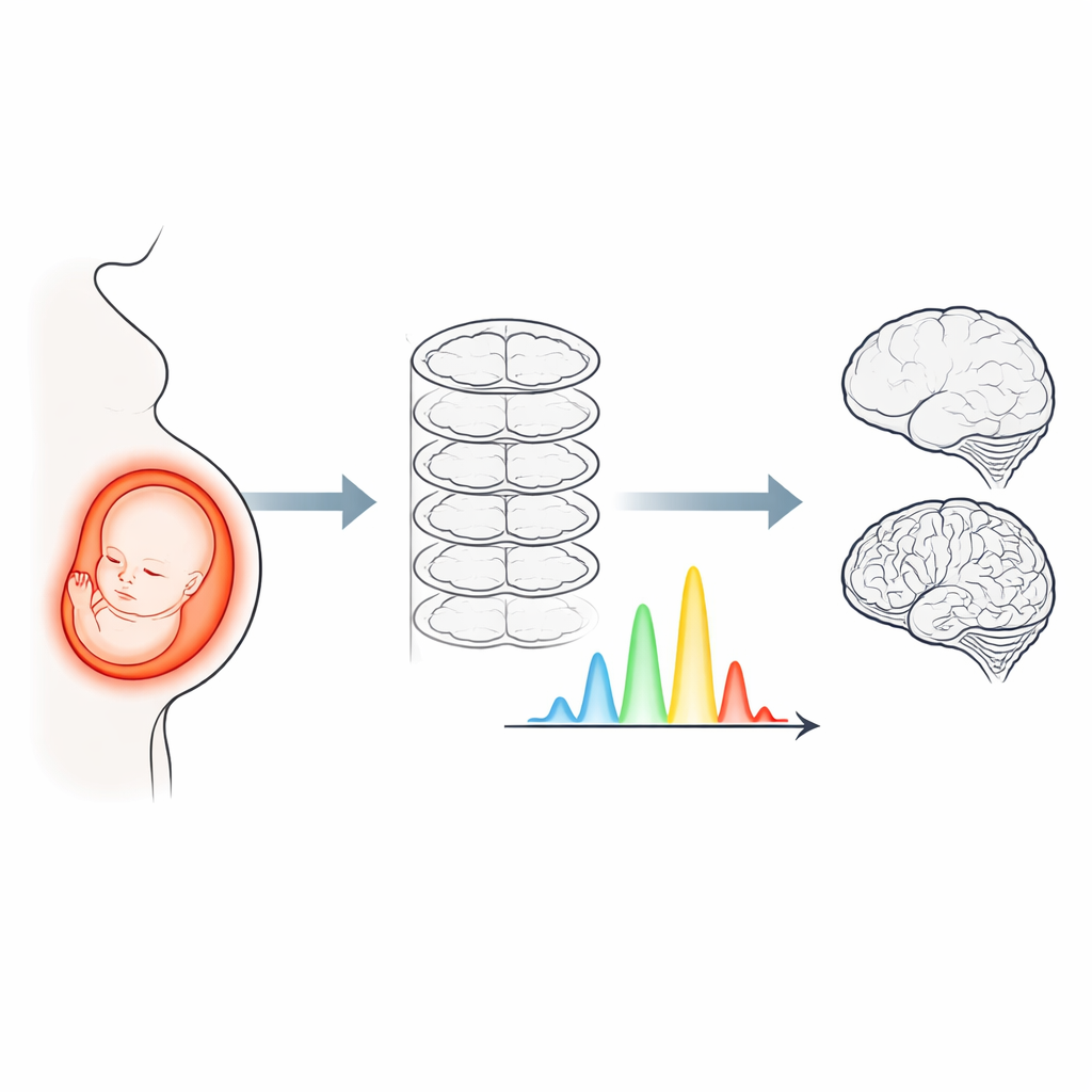
Watching Brain Folds Grow Before Birth
During pregnancy, the smooth surface of the fetal brain gradually transforms into a complex landscape of folds. This process, called gyrification, follows a fairly predictable schedule, with large folds appearing first and finer details emerging closer to birth and into early life. Clinicians currently judge whether this process is on track by visually inspecting ultrasound or MRI images. However, such assessments are subjective and can miss subtle or early changes, especially in conditions like lissencephaly, where the brain looks unusually smooth, and polymicrogyria, where the surface has many small, irregular folds.
Turning Brain Shape into a Signal
The researchers developed a method that treats the outline of each cerebral hemisphere in MRI images as if it were a signal that can be broken into different spatial “frequencies”—a bit like decomposing a sound into its bass and treble components. They extracted the outer contour of the brain from standard coronal MRI slices, converted these contours into a circular coordinate system, and then applied a mathematical tool called the Fourier transform. This produced a spectral profile for each fetus, summarizing how much of the contour’s shape is explained by broad, gentle curves (low frequencies) versus finer, intricate folds (high frequencies). From these profiles, they computed five overall measures, such as total power and how spread-out or skewed the spectrum was, along with the strength of the first twelve frequency components.
Typical Folding Patterns Across Pregnancy
The team analyzed MRI scans from 73 fetuses with typical development between 25 and nearly 38 weeks of gestation. In these fetuses, most spectral measures increased with gestational age, indicating that the brain surface becomes progressively more complex. Low-frequency components rose rapidly between about 24 and 32 weeks and then leveled off, matching the timing of early, large-scale folds. Mid-frequency components grew more steadily, while the highest frequencies surged later in pregnancy, echoing the emergence of finer folds. One early low-frequency component actually decreased over time, likely reflecting the shift from a simple, smooth, oval shape toward a more indented and lobed brain as key clefts such as the Sylvian fissure deepen.
Spotting Abnormal Folding in Rare Brain Conditions
Next, the researchers compared these typical patterns with spectra from 10 fetuses with lissencephaly and 14 with polymicrogyria. To ensure that differences were not simply due to being scanned at different weeks of pregnancy, they mathematically removed the effect of gestational age before comparing groups. Both malformation groups showed reduced total spectral power and lower “entropy,” meaning that their folding energy was less evenly distributed across frequencies. Lissencephalic brains had especially strong reductions in many frequencies, particularly those linked to large-scale features such as the Sylvian fissure, and showed a spectrum shifted toward low frequencies, consistent with a smoother, less varied surface.
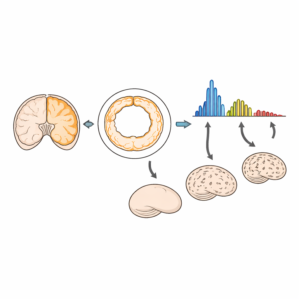
Surprising Insights into Many-Small-Folds Brains
Polymicrogyria, where the brain surface appears to have too many folds, might intuitively seem like it should increase high-frequency power. Instead, the spectral analysis revealed lower overall power and reduced contributions from several key frequencies. The authors suggest that this is because the additional folds in polymicrogyria tend to be shallow and irregular. In their framework, deeper folds contribute more strongly to the spectrum, so a brain with many small, thin folds can still show a net reduction in spectral power. Despite the complexity and variability of polymicrogyria, the method consistently detected abnormalities and even distinguished these cases from lissencephaly through differences in how spectral power was distributed.
What This Means for Future Pregnancies
By turning the brain’s outline into a spectrum of frequencies, this work offers a quantitative “fingerprint” of fetal brain folding that tracks normal maturation and flags departures from it. The approach works on standard 2D MRI scans, avoiding the need for time-consuming 3D reconstructions, and was sensitive to both globally smooth brains and those with many shallow, irregular folds. For parents and clinicians, such tools could eventually support earlier and more reliable diagnosis of cortical malformations, guiding counseling, follow-up imaging, and postnatal care. While larger and prospective studies are still needed, spectral analysis shows promise as a robust biomarker of how the fetal brain’s surface architecture develops—and when it starts to go off course.
Citation: Yehuda, B., Gal, R., Wexler, Y. et al. MRI-based spectral analysis of fetal brain gyrification in typical development and in lissencephaly and polymicrogyria. Sci Rep 16, 10018 (2026). https://doi.org/10.1038/s41598-026-38229-9
Keywords: fetal brain development, cortical folding, fetal MRI, lissencephaly, polymicrogyria