Clear Sky Science · en
Spatial localization of avian and human influenza A virus receptors in male and female bovine reproductive tissues
Why this matters for cows, farmers, and people
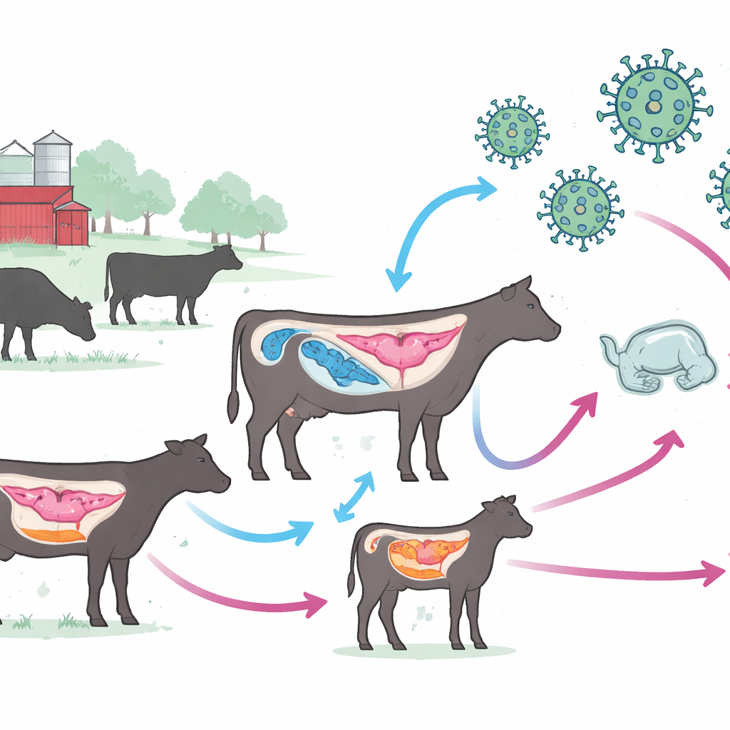
Highly pathogenic bird flu (H5N1) has recently jumped into U.S. dairy herds, with virus found in milk and mammary tissue. That raises an urgent question: could the virus also use the cow’s reproductive system as a hidden highway, spreading through semen, birth fluids, or even to developing calves—and potentially back to people? This study maps, in detail, where influenza-friendly docking sites are located in male and female bovine reproductive organs, offering an early warning about new routes of infection and spread.
How flu finds a foothold in the body
Influenza viruses begin infection by grabbing onto tiny sugar tips, called sialic acids, that decorate the surface of many cells. Bird-adapted strains, like classic H5N1, prefer one type of linkage; human-adapted strains favor another. Think of them as different keys that fit slightly different locks. The researchers used special plant proteins, known as lectins, that glow blue wherever these “locks” appear in tissue slices from bulls and cows collected at a slaughterhouse. By comparing staining patterns across organs, they built a spatial map of where avian-like and human-like flu receptors sit in the reproductive tract.
Hidden entry points in the bull
In males, the team found an extensive network of influenza-friendly sites. The penile urethra carried both human-like and one avian-like receptor type in its surface layers, and all three receptor forms lined blood vessels and connective tissue inside the penis. Receptors were also common in the seminal vesicles, vas deferens, epididymis, and testes. Importantly, the epithelium of the vas deferens and epididymis—which handle sperm storage and transport—showed strong human-like receptor staining, while surrounding tissues carried avian-like forms. Mature sperm themselves did not display avian-type receptors but did show human-type receptors concentrated in the acrosome and midpiece, regions crucial for fertilization and motility. Together, these patterns suggest that several parts of the male tract could allow virus entry, systemic spread via blood, or contamination of semen, even though actual virus has not yet been proven in bull semen.
Vulnerable spots in the cow
In females, receptor distribution was more patchy but strategically located. The vagina and cervix carried both human-like and one avian-like receptor type on their luminal surfaces, echoing known hot spots for other sexually and vertically transmitted viruses. The oviduct, the site of fertilization and early embryo development, showed high levels of all three receptor types on its inner surface, with human-like and one avian-like type also present in blood vessels beneath. The ovary contained avian-like receptors in follicle-supporting granulosa cells and human-like receptors in the egg’s outer coat, hinting that eggs and early embryos could potentially bind influenza. As expected from recent outbreak data, the mammary gland was rich in avian-type receptors in milk and alveolar structures, and in human-type receptors in epithelium and connective tissue, consistent with the gland serving as a powerful amplifier and source of virus-laden milk.
Pregnancy and the risk to the next generation
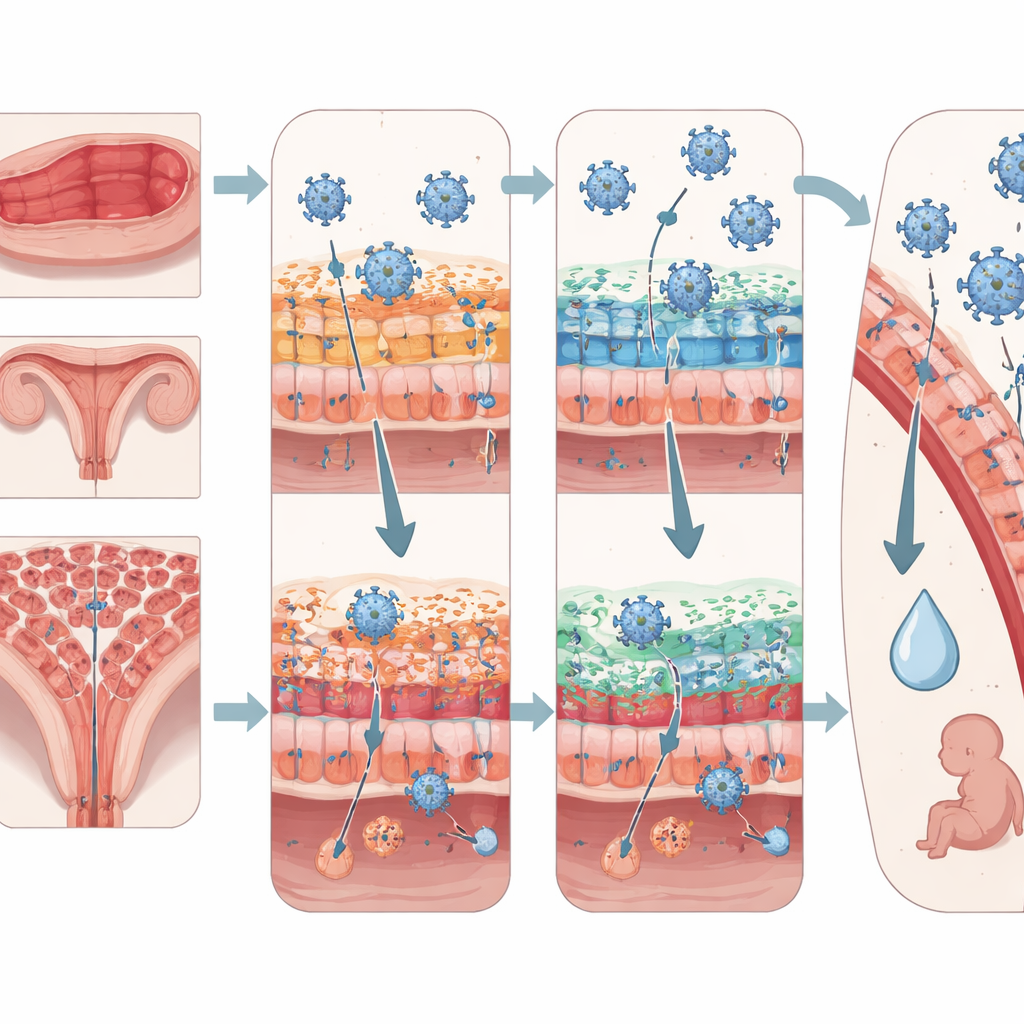
The uterus told a particularly important story. In non-pregnant cows, the inner lining that faces the uterine cavity showed little to no detectable flu receptors, even though blood vessels and underlying tissue carried human-like and one avian-like type. During early pregnancy, however, the picture changed dramatically: both human-like and avian-like receptors appeared along the luminal and glandular epithelium, as well as in the supporting stroma and muscle. Strikingly, receptor staining was also found on the conceptus itself—the early embryo and its surrounding membranes—on the surfaces that directly contact the uterine lining and in underlying endoderm. This configuration creates a potential bridge for virus to move between mother and developing offspring at a critical window for implantation and organ formation.
What this means for disease spread and control
By charting where influenza receptors sit in bovine reproductive tissues, this study shows that both bird-like and human-like flu strains have many potential docking sites in the cow’s body beyond the lungs and mammary gland. Male tissues appear broadly permissive along the semen pathway, while female tissues concentrate receptors in the vagina, cervix, oviduct, uterus during early pregnancy, ovary, and mammary gland. Although the work does not demonstrate active infection or transmission through these routes, it highlights plausible pathways for sexual, vertical, and milk-borne spread in cattle, and underscores the need for targeted biosecurity, surveillance of semen and reproductive tissues, and further experiments to test whether H5N1 and related viruses can truly exploit these newly mapped “ports of entry.”
Citation: Poliakiwski, B.D., Minela, T., Smith, D. . et al. Spatial localization of avian and human influenza A virus receptors in male and female bovine reproductive tissues. Sci Rep 16, 9974 (2026). https://doi.org/10.1038/s41598-026-36120-1
Keywords: H5N1 in cattle, bovine reproduction, influenza receptors, zoonotic transmission, avian influenza