Clear Sky Science · en
Application of simultaneous multi-slice accelerated readout-segmented echo planar diffusion-weighted imaging in assessing tumor response to neoadjuvant therapy in locally advanced rectal cancer
Why faster cancer scans matter
For people with advanced rectal cancer, radiation and chemotherapy before surgery can sometimes shrink the tumor so much that little or no cancer is left. In these cases, some patients may be able to avoid major surgery and its lifelong side effects. But this is only safe if doctors can tell, with high confidence, who still has cancer and who does not. This study tests a faster type of MRI scan to see whether it can reliably measure how well a rectal tumor has responded to treatment.
How rectal cancer is treated today
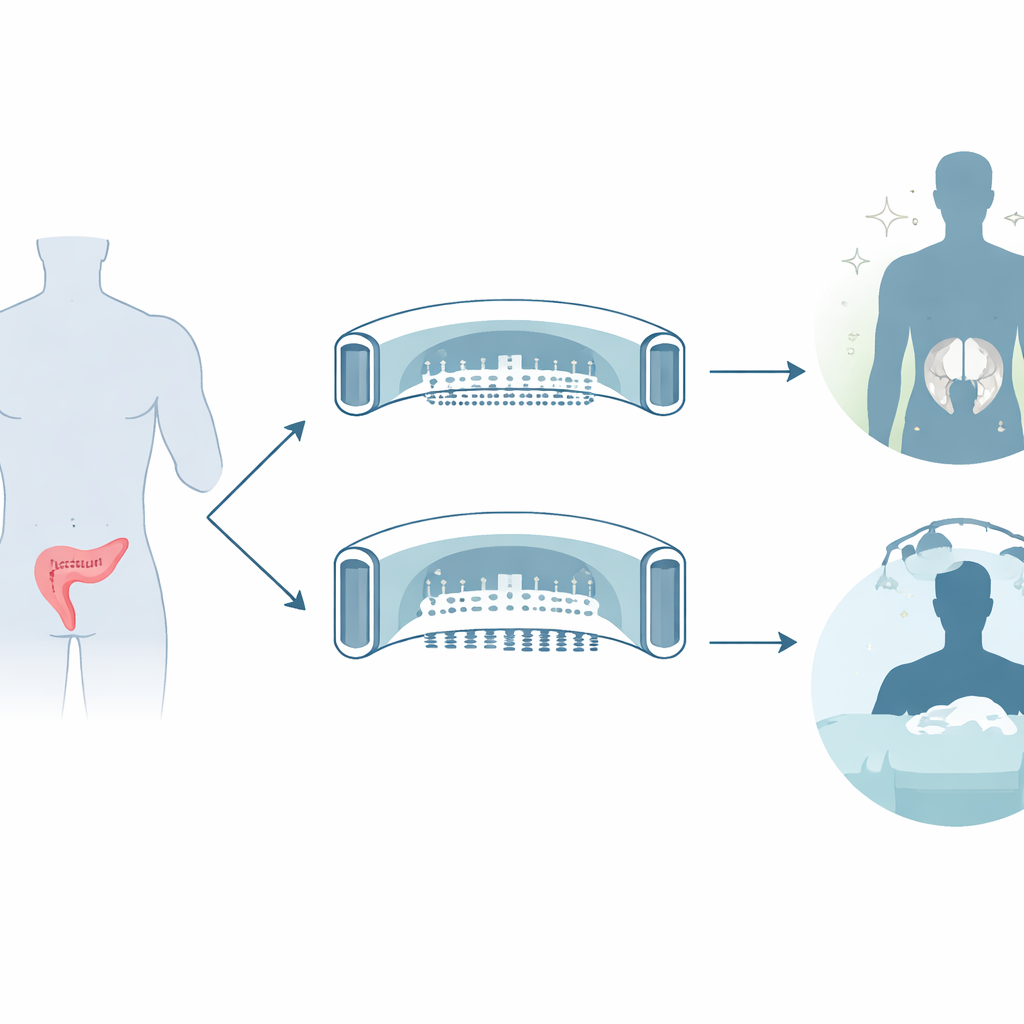
Standard care for locally advanced rectal cancer usually starts with a combination of chemotherapy and radiation, followed by surgery to remove the rectum and surrounding tissue. About one in five patients has a “complete response,” meaning no cancer cells are found when the tissue is examined under a microscope after surgery. Others have a strong but incomplete response, with only small areas of tumor left. If doctors could confidently identify these best responders before surgery, some might be offered a careful “watch-and-wait” approach or smaller operations that preserve more normal bowel function. Magnetic resonance imaging (MRI), especially when it tracks how water moves within tissue, is one of the main tools used to judge this response.
A new twist on MRI speed
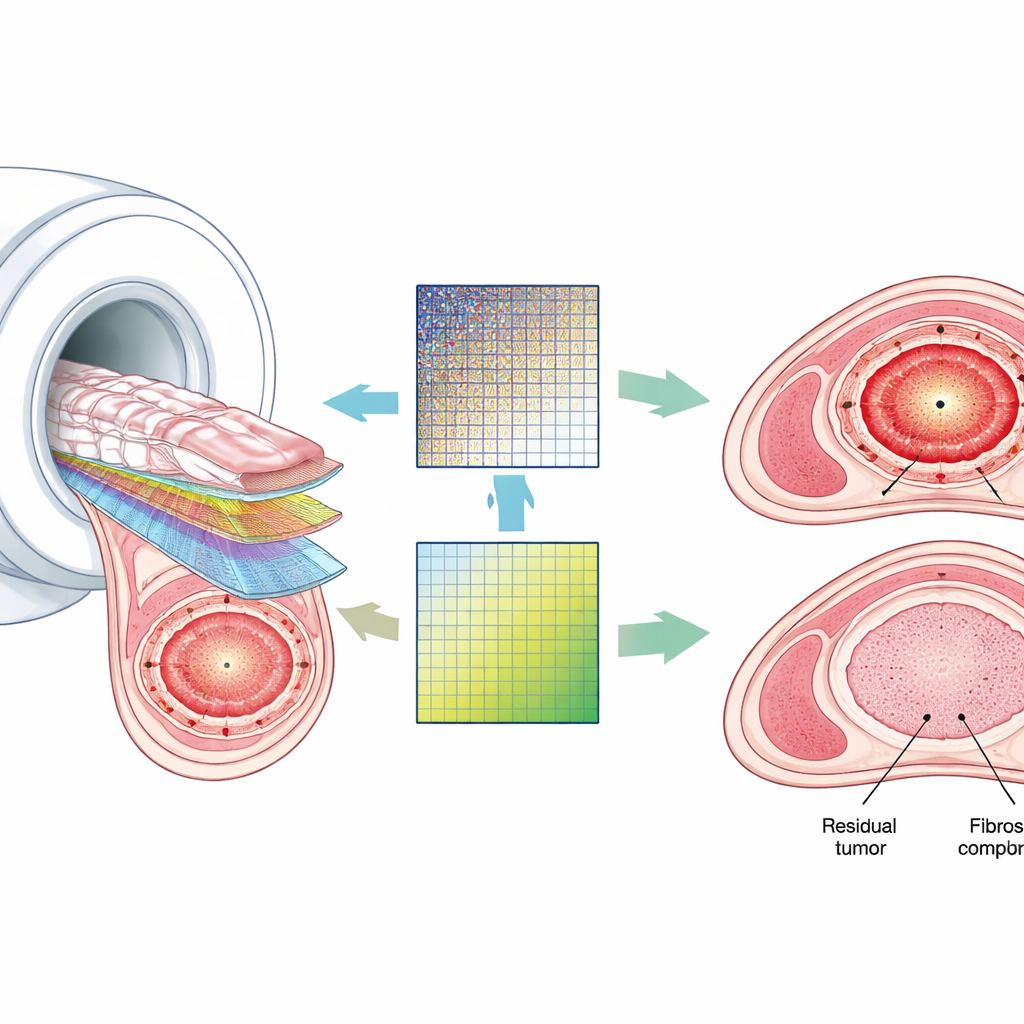
Conventional diffusion-weighted MRI of the rectum can produce detailed images that help distinguish dense tumor tissue from looser scar tissue by calculating a number called the apparent diffusion coefficient (ADC). However, the most accurate form of this scan, known as readout-segmented echo-planar imaging, takes several minutes to acquire and is prone to motion artifacts. The team tested a faster variant that captures several image slices at once, called simultaneous multi-slice RESOLVE. This approach nearly halves the scan time while keeping the same basic image design, raising the question of whether the quicker method can still match the slower one in judging treatment response.
What the researchers did
The study followed 42 patients with locally advanced rectal cancer who received chemotherapy and radiation before surgery. Each patient underwent a detailed rectal MRI including both the standard and the accelerated diffusion scans. Two experienced radiologists carefully outlined the treated tumor area on the images and recorded ADC values from both scan types, without knowing the surgical results. They also judged how much tumor appeared to remain and how deep it seemed to invade the bowel wall. After surgery, pathologists classified each case as a complete response or not, a good or poor response overall, and low or high tumor stage. The researchers then compared how well each MRI method’s ADC measurements could separate these groups.
What the scans revealed
In both the standard and fast scans, patients with a complete or good response had clearly higher ADC values than those with more residual tumor. Likewise, those whose tumors had been downstaged to a shallow depth after therapy showed higher ADC values than those with deeper invasion. Statistical analysis showed that the accelerated scan had diagnostic power similar to the conventional scan for identifying complete responders, good responders, and patients with lower post-treatment stage, even though the exact ADC numbers from the two methods were not interchangeable. Importantly, when the faster diffusion scan was added to routine MRI images, radiologists agreed with each other more often about tumor stage and response, and they were better at ruling out remaining cancer in patients who had in fact responded well.
What this could mean for patients
The findings suggest that a quicker diffusion MRI sequence can stand alongside the current, longer method for assessing how rectal tumors respond to pre-surgery treatment. While larger studies are needed, this faster scan may help radiology departments shorten exam times, reduce motion artifacts, and still provide the detailed tissue information needed to distinguish scar from active tumor. When combined with standard MRI pictures, it appears to improve confidence in identifying patients who likely have little or no cancer left after therapy. In the future, such imaging advances could support more personalized decisions—helping some patients safely avoid extensive surgery while ensuring that those who still harbor significant disease receive timely, aggressive treatment.
Citation: Yang, L., Zeng, Y., Hu, S. et al. Application of simultaneous multi-slice accelerated readout-segmented echo planar diffusion-weighted imaging in assessing tumor response to neoadjuvant therapy in locally advanced rectal cancer. Sci Rep 16, 11307 (2026). https://doi.org/10.1038/s41598-026-35617-z
Keywords: rectal cancer imaging, treatment response, diffusion MRI, neoadjuvant therapy, MRI techniques