Clear Sky Science · en
Single-cell chromatin accessibility landscape of cardiac non-myocytes identifies tissue repair program during heart regeneration
Why Hearts Heal Differently at Birth and Later in Life
Heart attacks and other serious cardiac injuries often leave permanent scars because adult hearts have very little ability to regrow lost tissue. Newborn mouse hearts, however, can briefly repair themselves almost perfectly. This study asks a deceptively simple question with big implications for future therapies: what are the support cells around heart muscle doing during this short window of natural regeneration, and how do they help decide whether the heart heals with healthy tissue or with scar?

The Hidden Helpers Around Heart Muscle
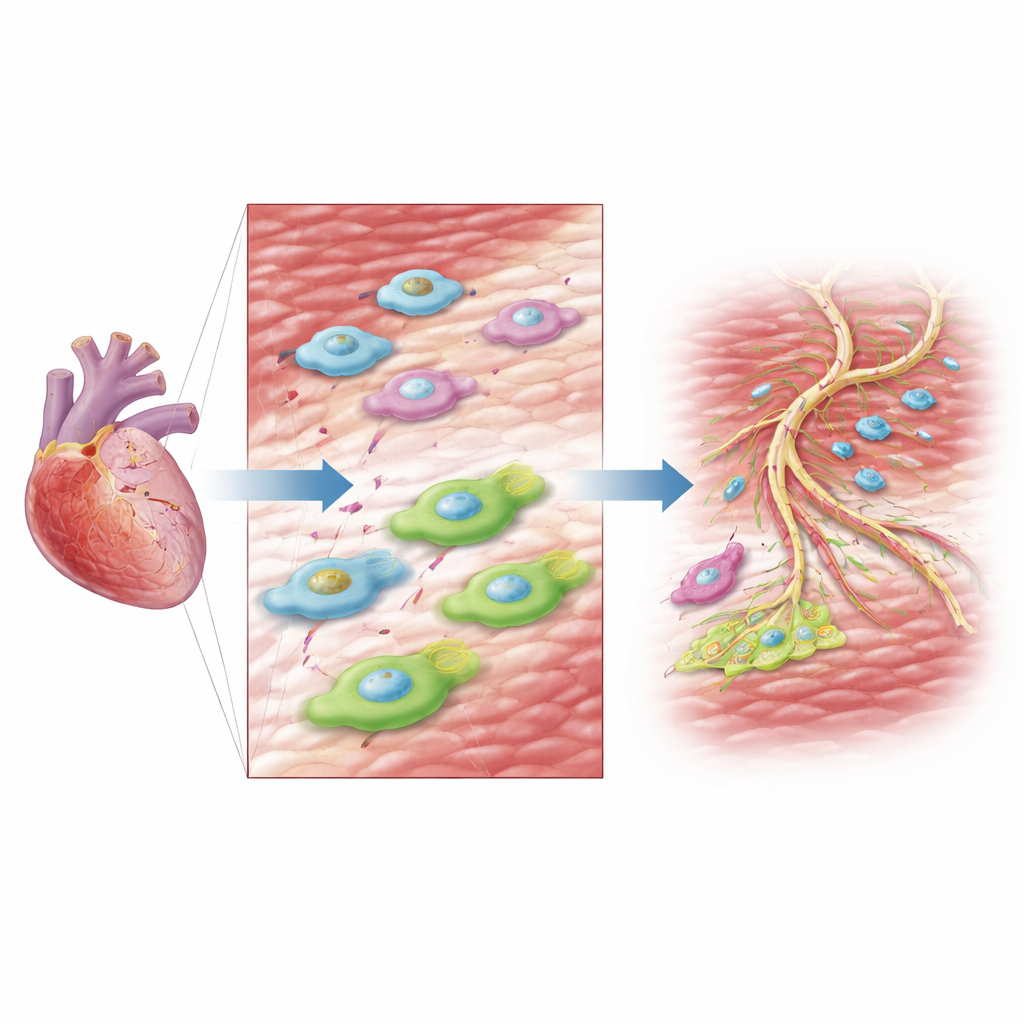
The heart is more than just beating muscle cells. It also contains a rich community of support cells, including fibroblasts that manage the tissue scaffold and endothelial cells that line blood vessels. The authors focused on these "non-muscle" cells in newborn mouse hearts that either could regenerate after injury or, in a genetically altered strain, failed to do so and healed with scar. Instead of measuring which genes were turned on, they used a technique that maps which regions of DNA are open and ready to be read in each single cell. This gave them an atlas of how the hearts’ support cells rewire their genetic control panels over time after injury.
A Short-Lived Repair Squad of Fibroblasts
Fibroblasts are often cast as the villains of heart disease because they can produce stiff scar tissue. Here, the team uncovered a more nuanced picture in newborns. They identified several fibroblast subgroups and found one special population that appeared only briefly after injury in hearts that regenerated well, and was almost absent in hearts that failed to regrow. These “regenerative fibroblasts” showed DNA regions open near genes involved in cell division, cell–cell contact, and tissue remodeling, suggesting a flexible, pro-repair state rather than runaway scarring. A transcription factor called CEBPD emerged as a key switch: its binding sites were especially accessible in this subgroup, and its activity rose sharply after injury.
Turning a Gene Switch into Real Repair
To test whether CEBPD truly drives helpful fibroblast behavior, the researchers dialed it down in newborn mouse hearts using virus-delivered genetic tools. Fibroblasts with reduced CEBPD failed to fully activate after injury: they divided less, produced fewer repair-related proteins, and showed weaker signs of entering the pro-regenerative state. At the whole-organ level, these hearts pumped blood less effectively and developed larger scars compared with controls, even though their baseline heart function before injury was normal. This shows that a carefully timed burst of fibroblast activation—guided by CEBPD—is not a problem to be avoided, but a requirement for proper neonatal heart repair.
Blood Vessel Builders and the AP-1 Signal
Regrowing heart muscle also demands a renewed blood supply. Within the endothelial cell population, the scientists identified an injury-induced subgroup that dominated early after damage in regenerative hearts. Its DNA landscape pointed to genes involved in forming cell junctions and new vessels. Motif analysis of its open chromatin highlighted another family of transcription factors, called AP-1, as major regulators. When the team blocked AP-1 activity with a small molecule, human endothelial cells in culture divided more slowly, migrated poorly, and formed fewer vessel-like tubes. In newborn mice, temporarily inhibiting AP-1 after heart injury reduced new vessel formation, weakened heart function, and increased scarring, but had little effect on uninjured hearts.
What This Means for Future Heart Repair
Taken together, the study shows that successful neonatal heart regeneration depends on precisely tuned behavior of non-muscle support cells. Fibroblasts must briefly switch into a CEBPD-driven repair mode, and endothelial cells must engage an AP-1–guided angiogenesis program to rebuild the vascular network. By mapping these responses at the level of DNA accessibility in individual cells, the work points toward more targeted ways to coax the adult heart into a similar regenerative state. Instead of broadly suppressing fibroblasts or boosting blood vessels, future therapies might aim to recreate this balanced, time-limited repair program—reducing scarring while helping damaged hearts rebuild themselves.
Citation: Chen, Z., Nie, Y., Huang, L. et al. Single-cell chromatin accessibility landscape of cardiac non-myocytes identifies tissue repair program during heart regeneration. npj Regen Med 11, 18 (2026). https://doi.org/10.1038/s41536-026-00465-y
Keywords: heart regeneration, cardiac fibroblasts, endothelial cells, single-cell epigenomics, angiogenesis