Clear Sky Science · en
In situ architecture of plasmodesmata in Physcomitrium patens resolved by cryo-electron tomography
Tiny bridges that let plant cells talk
Plants may look still, but inside their tissues, cells are constantly exchanging signals and nutrients. This traffic has to cross sturdy cell walls, which raises a puzzle: how do neighboring cells stay connected without leaving big gaps in their walls? This study zooms in on the tiny channels that solve this problem in a moss and reveals how their shape and internal hardware control when cells stay in touch and when they shut the door.
Hidden doorways in the plant wall
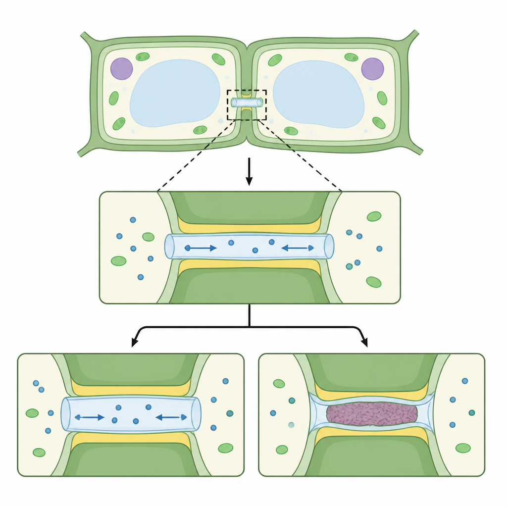
Plant cells are linked by microscopic tunnels that thread through their shared walls, creating direct bridges between the fluid and membranes of one cell and the next. In the moss Physcomitrium patens, the authors used a cryogenic imaging method that freezes tissue so fast that water does not form ice crystals. They then collected three dimensional views of these bridges inside intact tissues. The images show a simple but striking layout: each channel is lined by the outer cell membrane and holds a thinner inner tube that comes from the cell’s internal membrane network. The narrow space between outer wall and inner tube forms a sleeve where molecules can move from cell to cell, but its width varies along the channel and is tightest near the openings at each side.
How plants widen or seal the passages
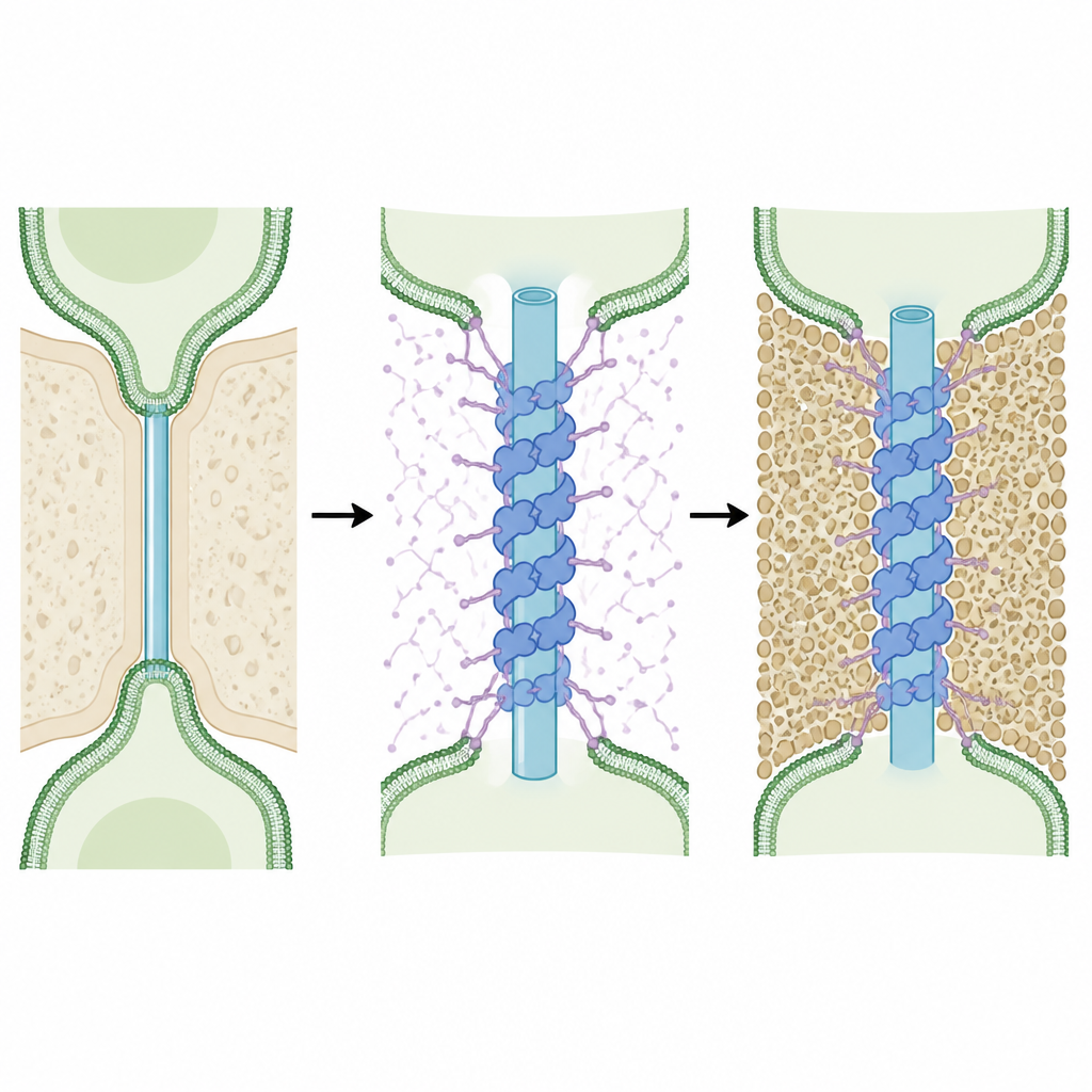
Plants adjust how easily molecules pass through these bridges, and this work links that control to changes in the surrounding wall material. The team examined three situations in moss filaments: normal tissue, tissue treated with the stress hormone abscisic acid and plants engineered to overproduce an enzyme that removes a wall polymer called callose. When abscisic acid was added, bulky, grainy deposits formed around the necks of the channels. In many cases these deposits completely pinched off the connection so that the inner tube, outer membrane and fluid sleeve were cut away from both cells and left buried in the wall. In contrast, when callose was actively removed, the channels became shorter and wider along their length. These changes match physical models that predict that wider, shorter tunnels should let molecules flow more easily, explaining why these modified plants show stronger cell to cell exchange.
A protein scaffold inside the channel
High resolution analysis of the inner tube revealed a surprising internal skeleton. Near the necks of the channels, the tube is wrapped by repeating rings of protein that wind around it in a helical lattice like the coils of a spring. These structures appear in both main moss tissues and under all tested conditions, marking them as core components of the channel design. By comparing the measured shapes with computer predicted structures of candidate proteins enriched at these bridges, the authors identified a family called Multiple C2 Domain and Transmembrane Proteins as the best match. Their models suggest that pairs of these proteins dimerize and pack together to form the helical coat, with one end anchored in the tube membrane and several compact domains interlocking to stabilize the assembly.
Flexible strands that shape traffic
Each protein in this family also carries a long, floppy segment that connects one of its domains to the rest of the molecule. Predictions and image analysis indicate that these flexible linkers can reach outward from the coated tube into the surrounding sleeve and toward the outer membrane. The authors propose that many such linkers act together as tethers that hold the inner tube in place, keeping it from collapsing or snapping during growth and during sealing events. Because these segments are rich in both positive and negative charges and are predicted to remain disordered, they may also fill the sleeve as a loose, dynamic mesh that influences which molecules can slip through, not only by size but also by charge. In this way, the wall polymer callose, which sets the sleeve width, and the protein linkers, which fill that space, could work together to fine tune the connectivity between cells.
Why these tiny bridges matter
This study delivers a detailed picture of how plant cell bridges are built and how they respond when a moss enters stress related states. It shows that a hormone signal can drive local remodeling of the wall that fully seals channels, while a specific protein scaffold and its flexible arms maintain the structure of the inner tube and help set the rules for molecular passage. For a non specialist, the key message is that plant cells are joined by highly organized, adjustable doorways whose architecture is central to how tissues grow, share resources and react to a changing environment.
Citation: Dickmanns, M., Pöge, M., Xu, P. et al. In situ architecture of plasmodesmata in Physcomitrium patens resolved by cryo-electron tomography. Nat. Plants 12, 1051–1061 (2026). https://doi.org/10.1038/s41477-026-02294-9
Keywords: plasmodesmata, plant cell communication, cryo electron tomography, cell wall, protein scaffolds