Clear Sky Science · en
Tuft cells shape airway remodeling by eliciting OXGR1- and SOX9-dependent stem cell programs
Why this airway repair story matters
Every breath brushes past a delicate lining that keeps germs, dust, and pollution at bay. When this barrier is damaged, our lungs and sinuses must quickly patch the leak. This study reveals that a rare type of cell, called a tuft cell, does more than just sense trouble. It can redirect how the airway repairs itself in ways that may harden tissues and weaken the barrier, shedding light on chronic conditions such as asthma and nasal polyps.
Guard cells that sense danger
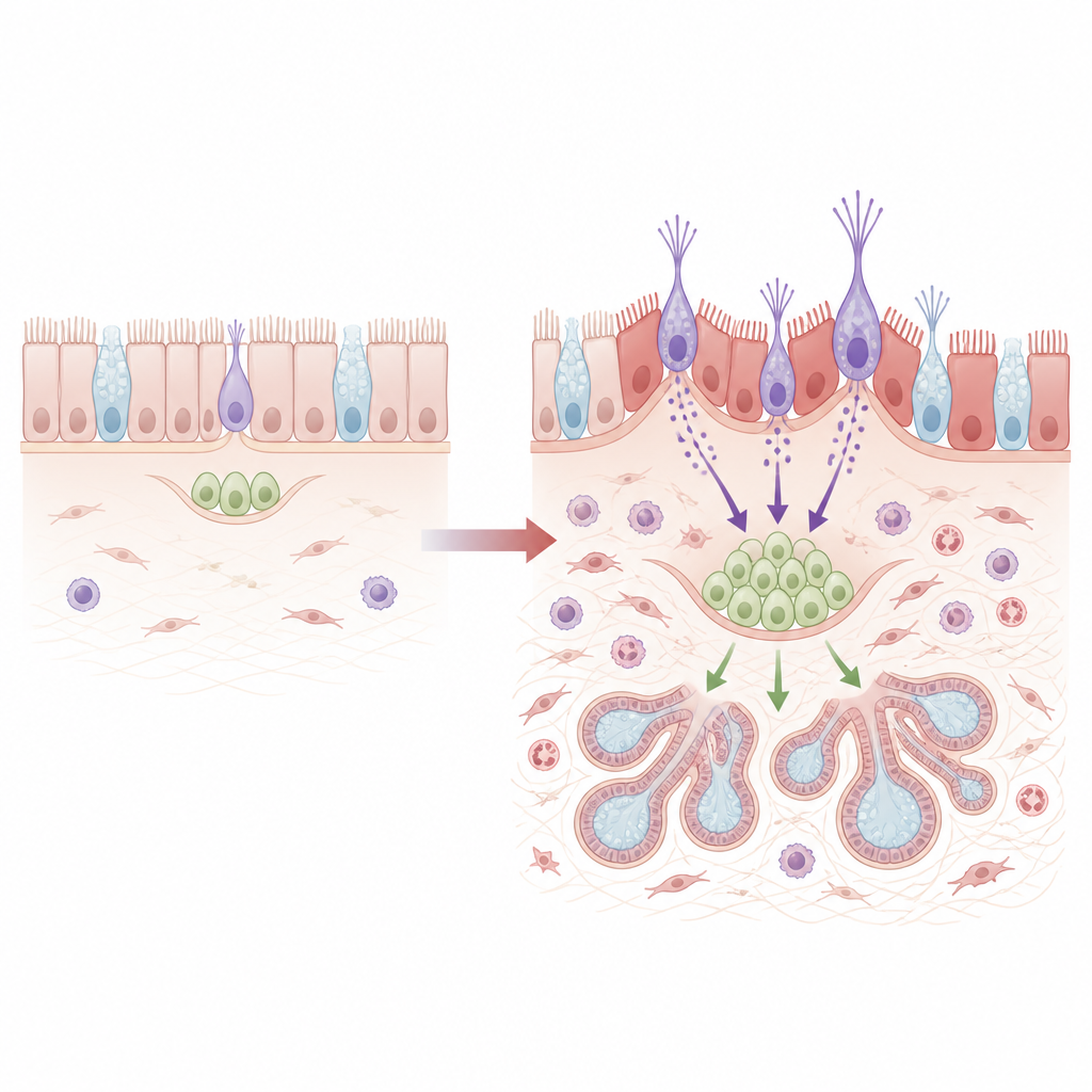
Tuft cells sit sparsely among the usual airway lining, but they are powerful chemical sentinels. They can detect invading microbes and irritating particles, then release bursts of signaling molecules that call in immune cells and trigger mucus release. The researchers used mice exposed to a mold allergen and to influenza virus to mimic repeated airway injury. They noticed that tuft cells multiplied under these stresses and asked whether these sentinels merely reported damage or actively guided how the tissue healed.
When repair goes off track
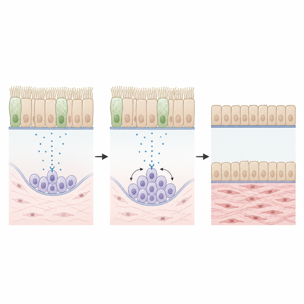
To track repair, the team compared normal mice with mice that lacked tuft cells or key tuft cell products. All groups initially suffered similar damage to their airway lining. Over time, though, clear differences emerged. Normal mice showed lingering DNA damage, more dying cells, and a leakier barrier that let tracer molecules seep below the surface. Mice without tuft cells, or without tuft cell production of a specific group of fat-based signals called cysteinyl leukotrienes, rebuilt a tighter, more intact lining. This suggested that tuft cell signals can actually hold back ideal repair and keep the barrier fragile.
Stem cells from deeper glands join the repair crew
Below the surface lining lie submucosal glands, which make mucus and harbor stem-like cells marked by a protein called SOX9. These cells can act as backup repair crews when damage is severe. By labeling these gland cells in advance, the scientists showed that, after injury, many of the surface stem cells in normal mice were in fact newcomers that had migrated up from the glands. This migration depended on tuft cells and their leukotriene signals acting through a receptor called OXGR1 on the gland cells. When tuft cells, leukotriene production, or OXGR1 were removed, far fewer gland-derived stem cells joined the surface, and the airway remodeled less, with fewer enlarged glands and less collagen buildup.
How the wrong repair program stiffens the airway
Gland-derived surface stem cells did not behave like the usual local stem cells. When the team isolated and sequenced their genes, these cells showed higher activity in switches linked to inflammation and to a shift toward more flexible, muscle-like traits. They also turned on many collagen genes and were poor at generating cilia, the tiny beating hairs that help clear mucus. In living mice, this matched a surface lined with fewer ciliated cells, weaker junction proteins that hold cells together, and more markers of stiff, fibrous tissue. Strikingly, when the researchers deleted SOX9 in airway stem cells, the lining regained abundant cilia, stronger cell-to-cell contacts, and less scarring, even after repeated injury.
Links to chronic sinus disease in people
To test whether this circuit matters in human disease, the team examined sinus tissue from patients with chronic rhinosinusitis with nasal polyps and from controls. Patients with polyps had many more tuft cells and more cells carrying both SOX9 and a muscle-like protein, along with reduced levels of a key barrier molecule called E-cadherin. The worse these changes were, the weaker the barrier signal appeared. Single-cell gene data from human basal cells showed an imprint of the same SOX9-driven program seen in mice, suggesting that this tuft cell–stem cell pathway is active in human airway remodeling.
What this means for future treatments
These findings outline a chain of events in which tuft cells sense airway damage, release leukotrienes, and activate OXGR1 on gland stem cells, which then move to the surface and rebuild it with a more fibrotic, less ciliated lining. While this backup system closes wounds, it leaves behind thickened tissue and a leakier barrier that may underlie chronic airway diseases. Targeting steps in this circuit, such as tuft cell leukotrienes, OXGR1, or SOX9 activity in stem cells, could help shift repair back toward a healthier, more flexible airway surface.
Citation: Lee, M., Wang, X., Ye, Q. et al. Tuft cells shape airway remodeling by eliciting OXGR1- and SOX9-dependent stem cell programs. Nat Commun 17, 4356 (2026). https://doi.org/10.1038/s41467-026-70763-y
Keywords: airway remodeling, tuft cells, stem cells, nasal polyps, lung repair