Clear Sky Science · en
Single-cell and spatial profiling reveal cDC2A-CXCL13+CD8+ T-epithelial cell crosstalk and cytotoxicity through TNFRSF9 in cutaneous and mucosal lichen planus
Why this skin disease matters
Lichen planus is a chronic condition that causes itchy, sometimes painful rashes on the skin and fragile sores in the mouth and genitals. For patients, it can be disfiguring, uncomfortable, and stubbornly resistant to treatment. Doctors have long known that the body’s own immune system attacks the skin in lichen planus, but they have not understood exactly which cells are responsible or how they communicate. This study uses powerful new “single-cell” and spatial mapping tools to draw a detailed map of the immune battle inside lichen planus lesions and to point toward a new treatment target.
A closer look at lichen planus types
Lichen planus comes in several forms. Cutaneous lichen planus affects the skin; mucosal lichen planus affects moist surfaces such as the mouth; and lichen planopilaris mainly attacks hair follicles on the scalp. The researchers collected skin, scalp, and mouth biopsies from 28 people with these conditions and 18 healthy volunteers. They then profiled more than 220,000 individual cells, asking which genes each cell was using and where each cell sat within the tissue. This allowed them to catalog many cell types, from structural skin cells to a wide range of immune cells, and to compare how these cells differ between diseased and healthy tissue.
Immune cells on the attack
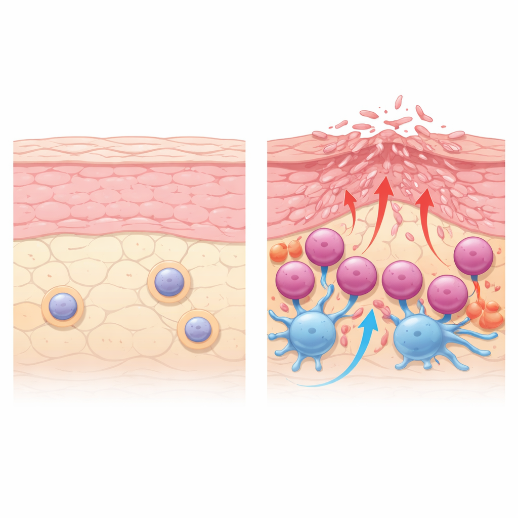
Across cutaneous and mucosal lichen planus, the team saw dense bands of immune cells hugging the boundary between the surface layer of the skin and the layer beneath it. In these regions, there was a strong signature of “killer” activity: genes used by cytotoxic T cells to punch holes in target cells and trigger their death were highly active. In contrast, signals related to another well-known inflammatory pathway, the IL-17 axis, were weak or absent. This suggests that tissue damage in lichen planus is driven mainly by direct killing of epithelial cells rather than by the type of inflammation seen in some other skin diseases. Lichen planopilaris looked different: it showed fewer invading T cells and less of this cytotoxic signature, hinting at a distinct underlying process.
A damaging dialog between key immune cells
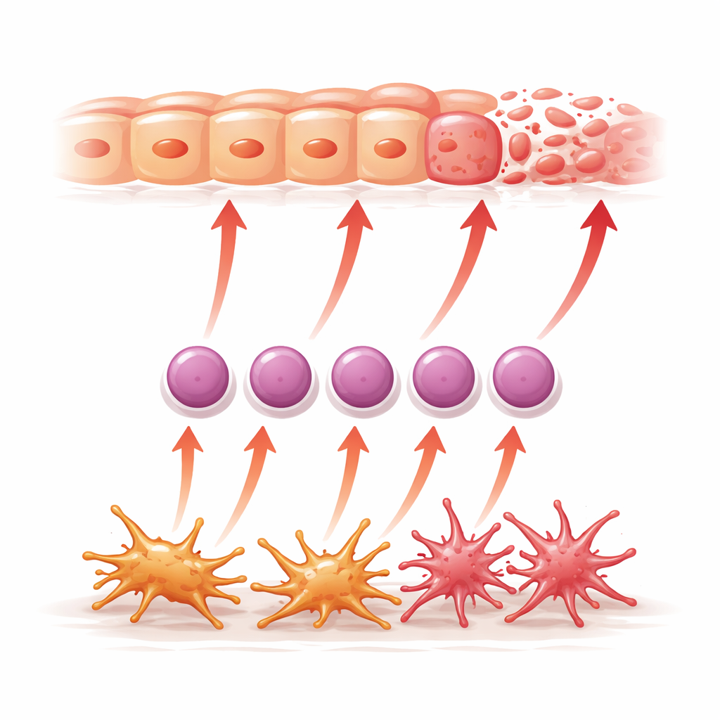
The study highlights two specialized immune cell types that appear to work together to drive disease in skin and mucosal lichen planus. One is a subset of dendritic cells, called cDC2A, that sit just below the surface layer. These cells produce the signaling molecule IL-15 and show strong activation of inflammatory pathways. Nearby is a distinct group of killer T cells that make the molecule CXCL13 and large amounts of interferon-gamma and other toxic factors. Spatial maps show these T cells clustering right against the epithelial layer they attack. The data suggest that IL-15 released by cDC2A cells helps push more ordinary CD8 T cells along a developmental path into highly cytotoxic CXCL13-producing cells that then damage the skin.
A switch that boosts T cell killing
As T cells mature from an early GZMK-rich state into the CXCL13-producing state, they ramp up genes linked to killing, including GZMB, IFNG, and TNFRSF9. TNFRSF9 encodes a surface receptor called 4-1BB that acts like a gas pedal for T cells when it binds its partner, 4-1BB ligand, on neighboring cells such as dendritic cells, fibroblasts, and epithelial cells. The authors detected clusters of 4-1BB–positive, granzyme-rich CD8 T cells precisely where tissue damage is most severe. In lab co-cultures of human skin cells and activated immune cells, blocking 4-1BB reduced death of the skin cells, showing that this pathway is not just present but functionally important. They also observed that patients treated with a JAK inhibitor, a drug that quiets certain immune signals, showed reduced TNFRSF9 in these T cells.
What this means for future treatment
By combining single-cell sequencing, spatial gene and protein mapping, and functional tests, this work paints lichen planus as a disease driven by a focused alliance between IL-15–producing dendritic cells and highly armed CXCL13-positive CD8 T cells that attack the skin and mucosal lining. The receptor 4-1BB emerges as a central amplifier of this attack, linking upstream signals from dendritic cells to downstream killing of epithelial cells. Because drugs that target 4-1BB are already being tested in cancer, this pathway represents a promising new avenue for therapies in cutaneous and mucosal lichen planus, with the potential to reduce tissue damage and improve quality of life for patients.
Citation: Jiang, R., Bogle, R., Xing, X. et al. Single-cell and spatial profiling reveal cDC2A-CXCL13+CD8+ T-epithelial cell crosstalk and cytotoxicity through TNFRSF9 in cutaneous and mucosal lichen planus. Nat Commun 17, 3962 (2026). https://doi.org/10.1038/s41467-026-70506-z
Keywords: lichen planus, single-cell sequencing, cytotoxic T cells, dendritic cells, immunotherapy targets