Why does our skin darken after sunlight or certain hormones, and how do pigment granules inside cells know when to turn on? This study uncovers an unexpected conversation between two tiny structures inside pigment cells—mitochondria, the cell’s “power plants,” and melanosomes, the compartments that make and store melanin. By watching these structures touch and separate in real time, the researchers reveal how these brief contacts help set up the right internal conditions for building melanin safely and efficiently.
Little Pigment Factories Inside Our Cells
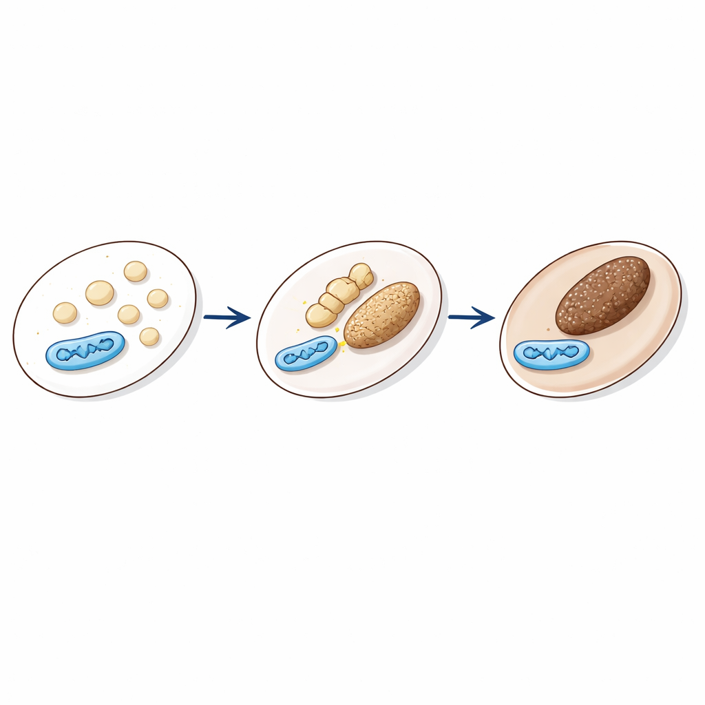
Melanin, the pigment that colors our skin, hair, and eyes, is made and stored in specialized compartments called melanosomes. These structures develop in stages, from pale, empty shells into dark, melanin-filled granules that can be sent toward the cell surface. Their activity is tuned by signals such as the hormone α-MSH, which rises after ultraviolet light exposure and boosts pigmentation. For melanosomes to work properly, the chemistry inside them—especially acidity and calcium levels—must change at the right time. One early step is the formation of a protein scaffold made of PMEL fibrils, which requires an acidic interior. Later, the compartment becomes less acidic so melanin-producing enzymes can function. How these precise changes are powered and timed has been unclear.
When Power Plants Meet Pigment Granules Figure 1.
The team focused on physical contact sites between mitochondria and melanosomes. Such organelle “handshakes” are known to be important elsewhere in cells, for instance between mitochondria and the endoplasmic reticulum. Here, the researchers engineered a live-cell reporter system called MiMSBiT that glows when mitochondria and melanosomes come close enough for two engineered protein fragments to reunite. Using this tool in mouse melanoma cells, they found that α-MSH and related signals caused a strong, but transient, increase in mitochondria–melanosome contacts. These contacts peaked about three hours after stimulation—the same window when melanosomes became most acidic and when PMEL fibrils were forming—hinting that the physical proximity of the two organelles is tightly linked to pigment granule maturation.
The Tethering Team: STIM1 and Mitofusin 2
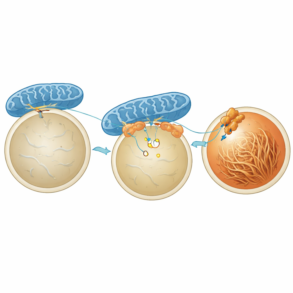
To understand what actually holds mitochondria and melanosomes together, the scientists homed in on a protein called MFN2, already known to help link mitochondria to other organelles. Knocking down MFN2 in pigment cells greatly reduced the hormone-triggered contacts and blunted the increase in pigmentation, without changing levels or basic activity of melanin-producing enzymes. The crucial player facing the melanosome turned out to be STIM1, better known as a calcium sensor in another cell compartment. Using a proximity-labeling method and high-resolution imaging, the researchers showed that a pool of STIM1 sits on melanosomes and briefly binds MFN2 on mitochondria when α-MSH is present. This interaction is triggered by a short-lived drop in calcium inside the melanosome lumen, which causes STIM1 to cluster and grab onto MFN2, forming a physical bridge.
Power Delivery and Acidification Inside Melanosomes Figure 2.
What is gained from pulling mitochondria and melanosomes together? The study shows that these contacts locally boost the availability of ATP, the energy currency of the cell, right at the melanosome surface. Using a fluorescent ATP sensor anchored at the melanosome membrane, the authors found that α-MSH raises ATP around melanosomes in a way that depends on mitochondrial energy production but not on sugar breakdown in the cytoplasm. When MFN2 or STIM1 were reduced, this local ATP surge disappeared, even though overall contact numbers or gross cell metabolism were not dramatically altered. The extra ATP appears to fuel proton pumps in the melanosome membrane that actively pump in protons, temporarily acidifying the interior. This acid pulse, in turn, promotes the assembly of PMEL into ordered fibrils that act as a scaffold on which melanin can later be safely deposited.
From Cell Contacts to Whole-Body Pigmentation
To test whether this microscopic mechanism matters in living animals, the researchers treated zebrafish embryos with a drug that interferes with the same MFN2 region needed for STIM1 binding. The developing fish showed markedly paler bodies, confirming that disrupting these organelle contacts impairs normal pigmentation in vivo. Together, the results outline a stepwise story: hormonal signals cause calcium changes within immature melanosomes; this activates STIM1, which teams up with MFN2 to tether mitochondria; those mitochondria then deliver ATP right where it is needed to acidify melanosomes and organize the PMEL scaffold; and only then can robust melanin production proceed. For a lay observer, this means that the color we see at the level of skin and hair depends on exquisitely timed, nanoscale interactions between tiny structures deep within each pigment cell.
Citation: Shiiba, I., Ishikawa, Y., Oshio, H. et al. STIM1-Mitofusin2 interactions tether mitochondria and melanosome contacts that promote melanosome maturation.
Nat Commun17, 3593 (2026). https://doi.org/10.1038/s41467-026-70282-w
Keywords: mitochondria–melanosome contact, melanin production, organelle communication, skin pigmentation, cellular energy and ATP