Clear Sky Science · en
Transcriptome analysis of the prefrontal cortex identifies inflammatory genes associated with cognitive impairment in a model of multiple sclerosis
Why brain inflammation matters for thinking
Many people with multiple sclerosis (MS) struggle not only with movement, but also with memory, concentration, and planning. These thinking problems can appear early and often progress over time, yet doctors still lack reliable ways to predict who will develop them or how to prevent them. This study zooms in on a key brain area for higher thought—the prefrontal cortex—to uncover how inflammation there may quietly erode mental abilities and point to new early warning markers. 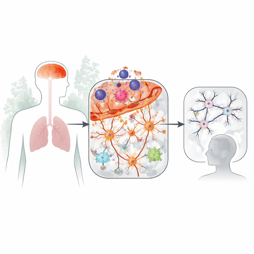
Looking inside a thinking hub of the brain
The prefrontal cortex helps us pay attention, juggle information, and make decisions. The researchers used a well-established mouse model of MS, called experimental autoimmune encephalomyelitis, in which the immune system attacks the brain and spinal cord. They focused on the acute phase of disease and extracted tissue from the animals’ prefrontal cortex. Using RNA sequencing, a technique that reads which genes are switched on or off, they created a global picture of how this thinking hub changes when inflammation is present.
Inflammation takes center stage
The analysis revealed that about 6% of all active genes in the prefrontal cortex changed during disease, and the vast majority were turned up rather than down. Genes linked to immune responses and inflammation dominated the scene, including those involved in antigen presentation (how cells show alarm signals to immune cells) and the complement system, a set of proteins that can tag and remove synapses. Many of the boosted genes are already known from human MS, strengthening the connection between this animal model and the human condition. In contrast, the genes that were dialed down were mostly tied to nerve cell communication and brain blood‑flow control, hinting that normal signaling is being dampened in this region.
Two levels of brain fire: low and high
When the team grouped the animals by their gene activity patterns, they found two clear subtypes: one with lower inflammatory activity (EAE-L) and one with much higher activity (EAE-H), even though the animals showed similar motor disability. In the low group, changes were mostly confined to the brain’s resident immune cells, microglia. In the high group, both microglia and star‑shaped support cells called astrocytes showed strong activation. Only in this high‑inflammation group did key neuronal and myelin‑related genes drop, including those important for synapses and for the insulating sheaths around nerve fibers. This suggests that as inflammation intensifies in the prefrontal cortex, it begins to directly disturb the cells and connections needed for healthy thinking. 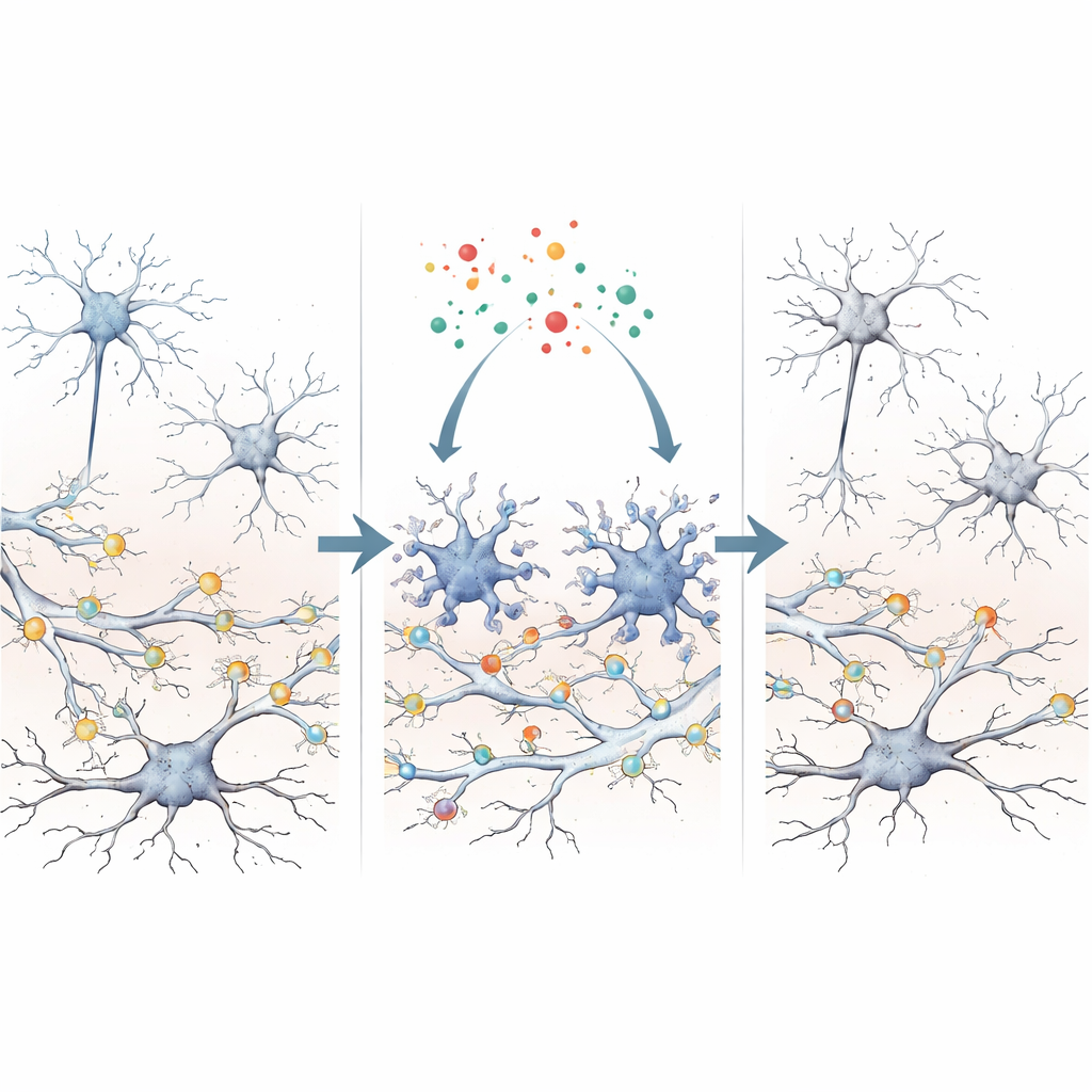
Linking molecular changes to memory problems
To connect these brain changes to behavior, the researchers tested mice on a task that measures recognition of object locations, a form of memory that relies on the prefrontal cortex. They assessed thinking ability before clear movement problems appeared, then later measured gene activity in the same brain region. Mice with poorer scores on the memory task tended to have higher levels of specific inflammatory genes, especially those encoding complement proteins known as C1q and molecules that drive antigen presentation. Other inflammatory genes, and genes directly linked to neurons, did not show this tight relationship. This pattern points to a particular inflammatory signature—rather than general sickness—as being closely tied to early cognitive decline.
From mice to patients: a promising fluid marker
Because C1q proteins can be measured in body fluids, the team tested cerebrospinal fluid from people with MS who either did or did not show clear cognitive impairment on formal testing. Despite having similar overall clinical profiles, patients with thinking problems had significantly higher levels of C1q in their spinal fluid. This finding mirrors the mouse data and suggests that an overactive complement system in the prefrontal cortex may help drive synapse loss and cognitive decline, and that C1q measurements could serve as a useful indicator of this hidden process.
What this means for people living with MS
Overall, the study shows that inflammation in the prefrontal cortex can be present and harmful even when outward disability looks similar, and that it is closely tied to early thinking problems. A specific group of inflammatory genes, particularly those related to the complement system and antigen presentation in microglia and astrocytes, stands out as a potential early warning signal. In the future, tracking markers like C1q in spinal fluid—and eventually, perhaps, in blood—could help identify people with MS who are at higher risk of cognitive decline and open the door to treatments aimed at calming this brain‑specific inflammation before it erodes the circuits that support memory and decision‑making.
Citation: Zupo, L., Adinolfi, A., Pieraccioli, M. et al. Transcriptome analysis of the prefrontal cortex identifies inflammatory genes associated with cognitive impairment in a model of multiple sclerosis. Cell Death Discov. 12, 177 (2026). https://doi.org/10.1038/s41420-026-03051-9
Keywords: multiple sclerosis, cognitive impairment, prefrontal cortex, neuroinflammation, complement system