Clear Sky Science · en
Directional dark field for nanoscale full-field transmission X-ray microscopy
Seeing Hidden Patterns in Everyday Materials
From the strength of a child’s tooth enamel to the durability of advanced composites, many important properties of materials are controlled by structures that are far too small to see with ordinary microscopes. This paper introduces a new X-ray imaging technique that can reveal not just the presence of tiny structures, but also the directions in which they are aligned—at length scales of tens of nanometers. That directional information is crucial for understanding how natural and man‑made materials are built, and how they fail.
Why Ordinary X-Ray Images Miss So Much
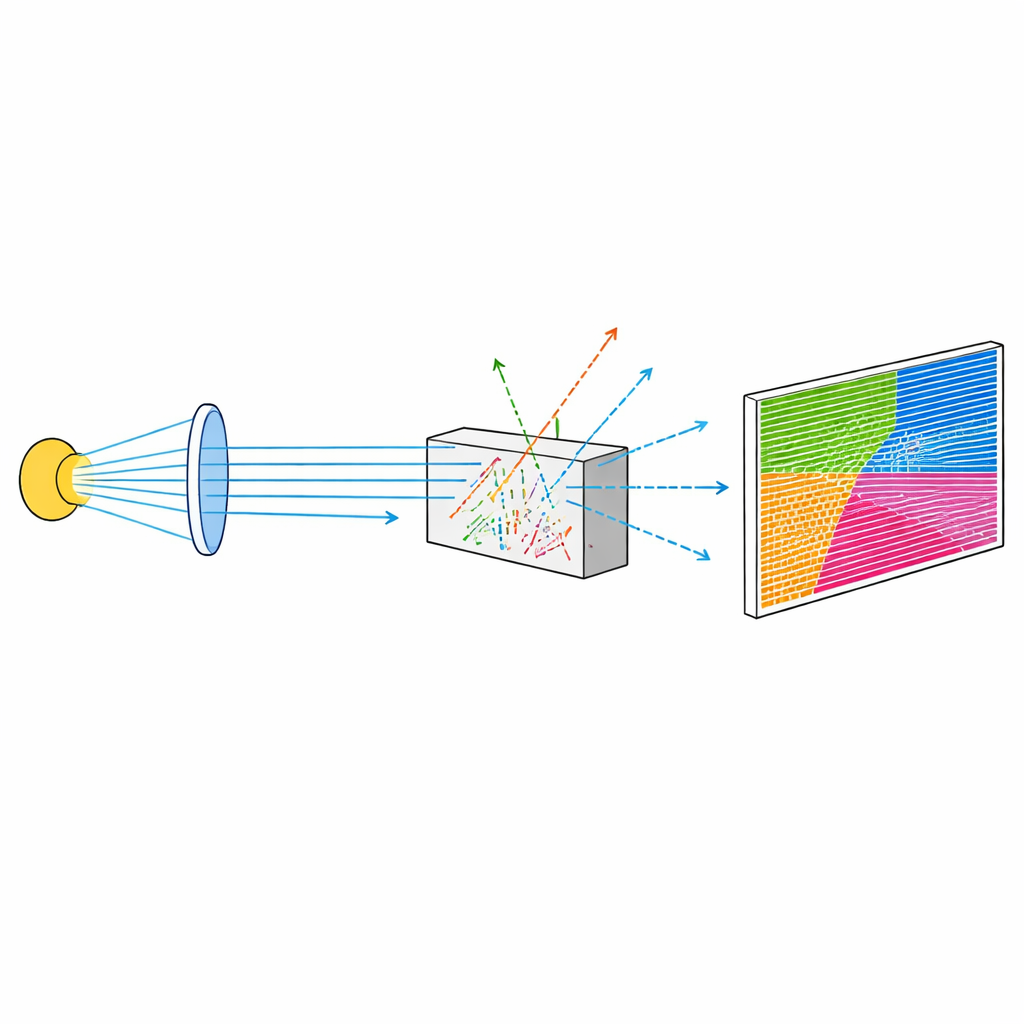
Conventional X-ray images mainly show how much of the beam is absorbed as it passes through a sample. This works well for bones or dense inclusions, but it struggles with subtle features such as fine pores, tiny cracks, or bundles of nanocrystals. To overcome this, researchers developed “dark‑field” X-ray imaging, which does not look at the direct beam but at X-rays that are scattered by small internal structures at very shallow angles. Dark-field images are exquisitely sensitive to inhomogeneities that remain invisible in standard attenuation or phase‑contrast pictures. Until recently, however, dark‑field methods that could tell which way structures were oriented were confined to micrometer scales and relatively coarse resolution.
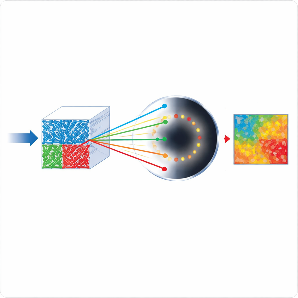
A New Way to Map Tiny Directions
The authors extend directional dark‑field imaging down to the nanoscale using a full‑field transmission X-ray microscope. They do this by adding movable apertures in front of the microscope’s condenser, which splits the X-ray beam into many small beamlets. By selectively blocking parts of the condenser, they allow only X-rays coming from specific directions to illuminate the sample. Scattered X-rays from these selected directions are then picked up in a region that is normally in the “shadow” of the microscope optics. By repeating the measurement with the condenser blocked from different sides and combining the results, the method reconstructs, for every image pixel, both how strong the scattering is and the preferred direction of the underlying structures—even when those structures are smaller than the pixel itself.
Testing with Tiny Patterns and Porous Pillars
To prove the concept, the team first imaged a gold test pattern shaped like a Siemens star and fine line pairs. In the directional dark‑field images, vertical and horizontal features lit up differently depending on which side of the condenser was used, clearly revealing the dependence of scattering on orientation. Remarkably, line pairs with features as small as 30–40 nanometers, well below the spatial resolution of the microscope, still produced a measurable directional signal. The method could even detect inconsistencies where some of these ultra‑fine lines had collapsed. Next, the researchers turned to a hierarchically nanoporous silicon pillar made by 3D printing an alloy and then selectively removing one component. The material contained large elongated pores made up of nanometer‑scale ligaments. The directional dark‑field projection revealed two main regions inside the pillar where the internal structure rotated by nearly 19 degrees. Independent phase‑contrast images of a slice through the same pillar confirmed a similar rotation, showing that the new approach can track subtle orientation changes in complex porous materials.
Looking Inside Defective Tooth Enamel
The technique was then applied to a pillar of enamel from a human tooth affected by molar incisor hypomineralization, a condition common in children. Enamel is built from long, thin crystals of hydroxyapatite bundled into rod‑like prisms. In the directional dark‑field image, the outer edges of these prisms appeared as fish‑scale structures whose orientations could be cleanly separated. Even more striking, the signal from within the prisms changed color across the sample, indicating that the average crystal direction rotated by more than 20 degrees between regions. This suggests that the method is sensitive to how the nanocrystals themselves are arranged inside each prism—information that is difficult to access by other means and may be important for understanding why diseased enamel is weaker. Solid support structures, by contrast, appeared dark, confirming that the image contrast really came from scattering by nanoscale features.
Pushing to Smaller Features with Smart Illumination
Beyond simply measuring directions, the authors show that they can tune which feature sizes contribute most strongly to the dark‑field signal. By exploiting the extra shadow region created when large parts of the condenser are blocked, they extend the range of detectable scattering angles. This effectively shifts the method’s sensitivity toward smaller structures. Experiments with a special “elbow” test pattern containing line pairs from 1000 down to 30 nanometers demonstrated that opening the dark‑field apertures into this extended shadow region boosts the signal from the smallest features, down to about 50 nanometers in the specific setup used. In principle, carefully designed illumination and apertures could make the technique selectively sensitive to chosen size ranges inside a complex material.
What This Means for Future Materials and Medicine
This work shows that directional dark‑field X-ray imaging can now map the orientation of structures tens of nanometers wide across relatively large fields of view, using a setup that can be added to existing transmission X-ray microscopes. It provides information beyond what standard dark‑field, attenuation, or phase‑contrast images can offer, and works for a range of samples from engineered nanoporous silicon to diseased tooth enamel. With brighter fourth‑generation synchrotron sources and improved optics, exposure times could shrink enough to follow changes in real time, for example as materials deform, crack, or undergo chemical reactions. Ultimately, this nanoscale “compass” for internal structure could become a powerful tool for designing better biomaterials, diagnosing subtle tissue changes, and optimizing advanced manufactured components.
Citation: Wirtensohn, S., Flenner, S., John, D. et al. Directional dark field for nanoscale full-field transmission X-ray microscopy. Light Sci Appl 15, 223 (2026). https://doi.org/10.1038/s41377-026-02263-z
Keywords: directional dark-field X-ray imaging, nanoscale transmission X-ray microscopy, nanostructure orientation, nanoporous materials, tooth enamel microstructure