Clear Sky Science · en
A “turn-off” photoacoustic contrast for urokinase-type plasminogen activator activity
Why tiny colon growths matter
Most colorectal cancers start as small growths called polyps in the lining of the colon. Doctors can see and remove many of these during colonoscopy, but very small or flat polyps are easy to miss and hard to judge as harmless or dangerous. This study describes a new type of imaging dye that could help reveal how aggressive a polyp is by sensing the activity of a cancer-related enzyme, giving doctors a clearer picture than size and shape alone.
A chemical clue to cancer aggressiveness
Colorectal cancer is one of the leading causes of cancer death worldwide, yet it often develops slowly over years. Current screening tools focus on finding and removing polyps, but about one in five are still missed, especially when they are less than five millimeters across. Even when detected, doctors cannot always tell which ones will turn into cancer. The authors focus on a protein called urokinase-type plasminogen activator, or uPA, which is more active in aggressive colon tumors and is linked to higher risk of spread and recurrence. Instead of measuring how much of this protein is present in tissue samples, they set out to image how active it is directly inside the body.
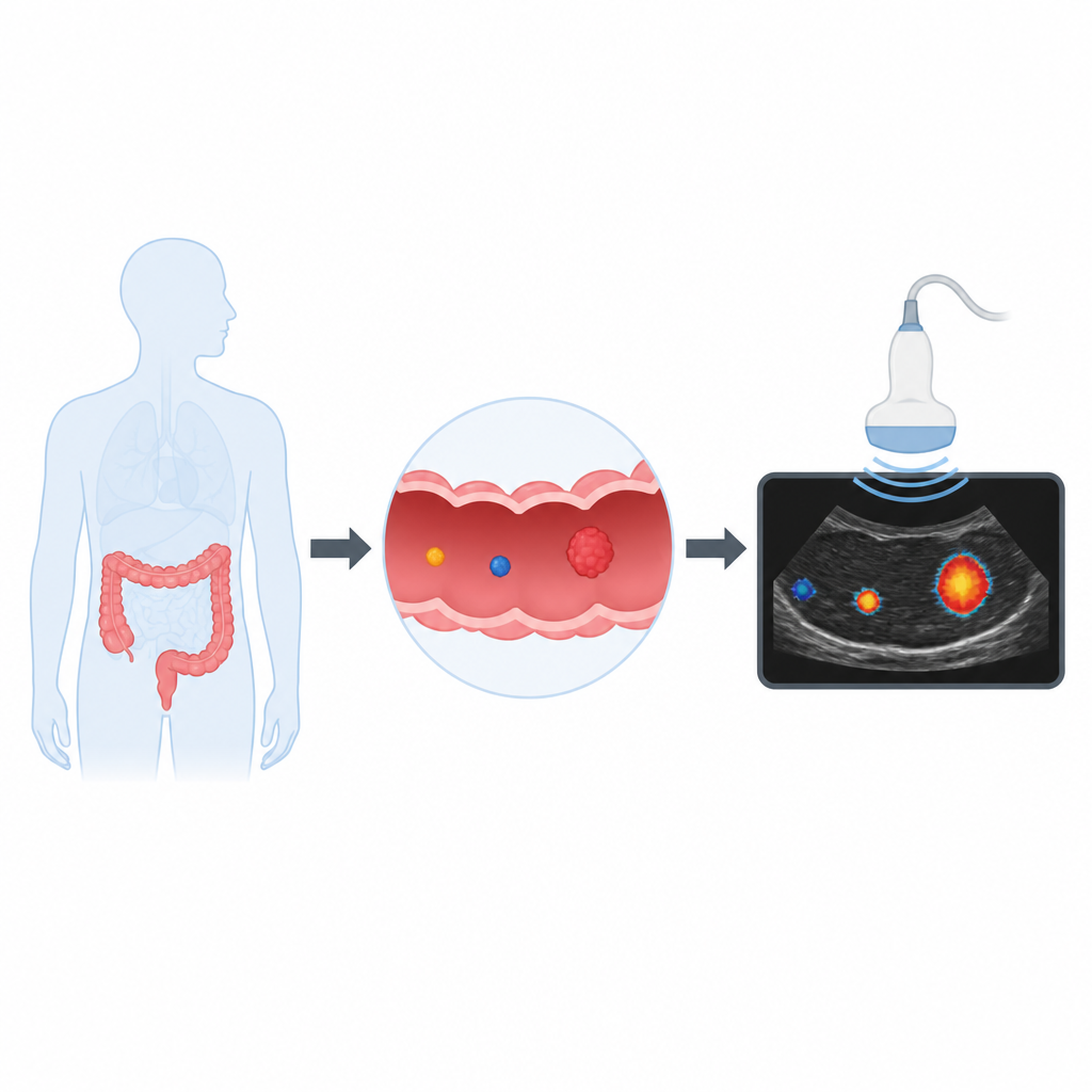
Turning sound into a picture
The team built their probe for a technique known as photoacoustic imaging. In this method, short pulses of near-infrared light are delivered into tissue. Special dyes absorb the light and heat up very slightly, causing tiny pressure waves that can be detected as ultrasound. This combines the chemical sensitivity of optical imaging with the depth and sharpness of ultrasound, and avoids radiation. The researchers attached a small three–amino acid piece, which uPA can cut, to a near-infrared dye. In its intact form, the dye produces a strong photoacoustic signal; when uPA cuts the peptide, the dye’s structure changes and its signal largely disappears. In other words, the probe starts “on” and then switches “off” in places where uPA is highly active.
Designing a smart, stable probe
To make this work in watery, biological environments, the scientists carefully tuned the chemistry of their dye and linker. They created a small molecule, called GGR-IR780, that dissolves well in water, absorbs light in a near-infrared range where the body’s own molecules produce little background, and gives a strong photoacoustic response at low concentrations. Laboratory tests showed that the dye is more stable than the starting dye material and keeps its signal over several hours of repeated light exposure. In gel-like phantoms that mimic tissue, the probe produced a clear, localized signal from a tube just 2.5 millimeters wide, suggesting it could pick up very small targets similar in size to tiny colon polyps.
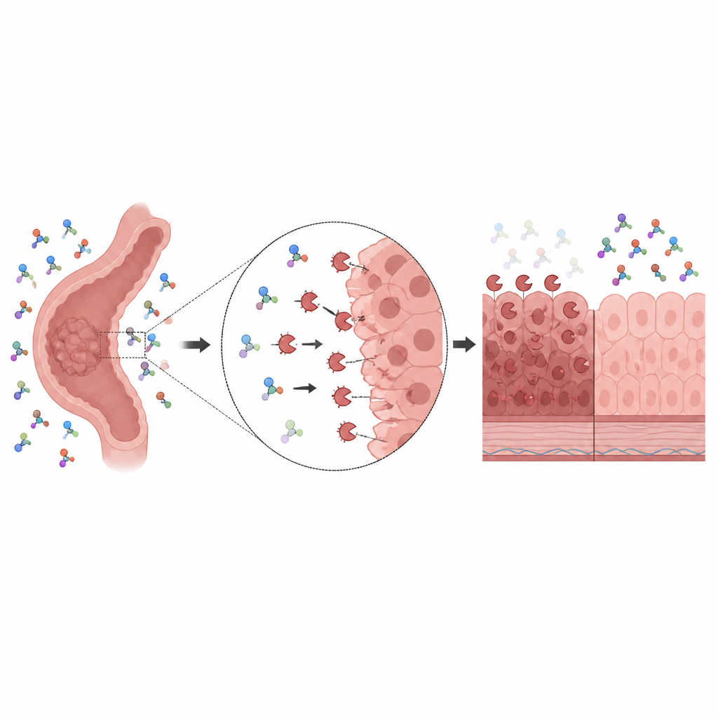
Watching the signal fade where enzymes are busy
Next, the authors asked how well the probe responds when uPA is present. In simple solutions, adding the enzyme caused the photoacoustic signal to drop by more than half within four hours, while ordinary fluorescence measurements took much longer to show a similar change. This indicates that photoacoustic imaging can track enzyme activity with higher temporal sensitivity. By varying enzyme and probe amounts, they also worked out how strongly uPA binds to the probe and how fast it acts on it, finding high binding affinity but only moderate speed of reaction. Studies using chromatography, mass spectrometry and nuclear magnetic resonance suggested that once uPA cuts the peptide, the dye fragment becomes unstable and tends to break down or clump, explaining why the signal vanishes rather than brightens.
Distinguishing aggressive from mild cancer cells
To test whether the probe can tell aggressive from less aggressive disease, the team turned to two human colorectal cancer cell lines. One, HCT-116, is known to invade and spread readily; the other, Caco-2, behaves more mildly. Protein tests and a standard color-based enzyme assay confirmed that HCT-116 cells have much higher uPA activity. When cell extracts from each line were mixed with the probe and imaged photoacoustically over time, the signal in the aggressive HCT-116 samples fell by about 56 percent within three hours, compared with only 33 percent for Caco-2. The probe also responded somewhat to another enzyme, cathepsin B, which is itself more abundant in aggressive cells. This partial cross-reactivity actually reinforces the ability of the probe to highlight more dangerous tumor types.
What this could mean for future colon screenings
Altogether, the study presents a small, water-soluble dye that acts as a “turn-off” beacon for regions with high activity of enzymes linked to aggressive colorectal cancer. While the work so far has been done in solutions, tissue phantoms and cell preparations, it shows that a strong initial photoacoustic signal followed by a local fade could be used as a real-time readout of how threatening a lesion is. With further testing in animals and, eventually, in humans, such probes might be delivered during endoscopy to mark polyps that deserve removal or closer follow-up, helping clinicians move from simply finding growths to quickly judging their true risk.
Citation: Sharma, A., Panda, S.K., Hasan, T. et al. A “turn-off” photoacoustic contrast for urokinase-type plasminogen activator activity. npj Biomed. Innov. 3, 33 (2026). https://doi.org/10.1038/s44385-026-00088-4
Keywords: colorectal cancer, photoacoustic imaging, molecular probe, urokinase activity, cancer aggressiveness