Clear Sky Science · en
Detection of α-synuclein aggregates in synucleinopathies: current approaches, biomarkers and challenges
Why this brain protein matters
Parkinson’s disease and related disorders quietly damage the brain for years before movement problems or memory loss appear. At the heart of these conditions is a small protein called alpha-synuclein that can clump into harmful aggregates. For decades, doctors could confirm these clumps only after death, by examining brain tissue. This review explains how scientists are now learning to detect these protein clumps in living people, using spinal fluid, tiny skin samples, scans, and even experimental nanosensors. These tools could help diagnose disease earlier, tell similar disorders apart, and track whether new treatments are working.
From hidden clumps to detectable signals
In Parkinson’s disease and dementia with Lewy bodies, alpha-synuclein aggregates inside nerve cells, while in multiple system atrophy it builds up in support cells. These deposits disrupt brain circuits, but they are hard to see directly in life. Researchers have therefore focused on biomarkers, measurable signs in fluids or tissues that reveal the presence of these clumps. A major advance is the use of “seed amplification” tests, which mimic the way a few abnormal protein clumps can trigger many more to form. By mixing a patient sample with normal alpha-synuclein in a test tube and shaking or sonicating it, scientists can coax any hidden seeds to grow into detectable fibers that bind fluorescent dyes.

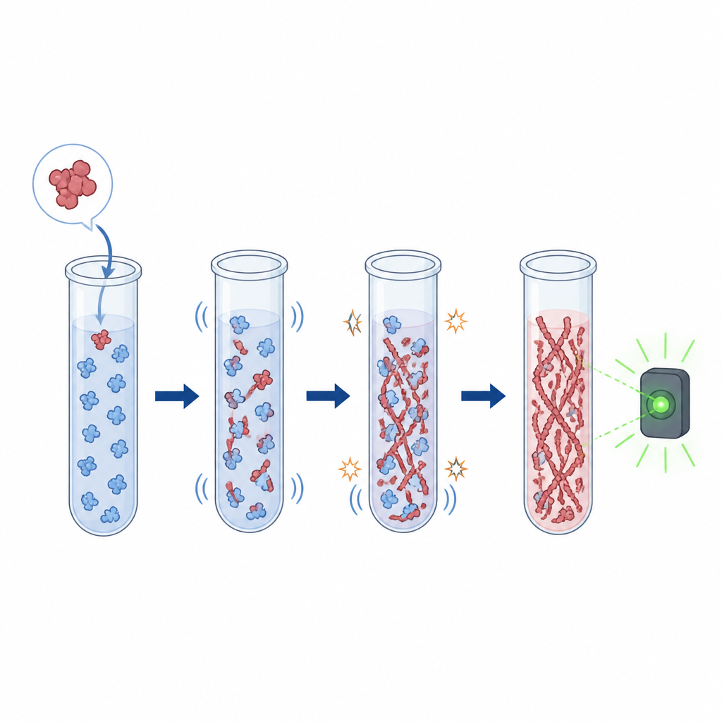
Powerful amplification tests for early detection
Several seed amplification methods have been developed, with real-time quaking-induced conversion, or RT-QuIC, emerging as the most widely validated. In this assay, shaking cycles drive the formation of new fibers if seeds are present, while a fluorescent signal tracks the reaction over time. Large studies show that RT-QuIC on spinal fluid can distinguish patients with Parkinson’s or dementia with Lewy bodies from healthy people with high sensitivity and specificity, and often becomes positive even in people who only have early warning signs such as sleep disturbance or loss of smell. Related methods, such as protein misfolding cyclic amplification and ultrasonication-based systems like HANABI, help reveal that different diseases harbor distinct “strains” of alpha-synuclein fibers, offering clues for telling Parkinson’s and multiple system atrophy apart.
Sampling beyond the brain
Because spinal taps are invasive, scientists are working to detect alpha-synuclein clumps in more accessible tissues. RT-QuIC and related assays now pick up seeds in samples from the nose lining, skin, salivary glands, gut, and blood, though performance varies with tissue site and disease stage. Traditional pathology tools are also being adapted: small skin biopsies can be stained to reveal abnormal, phosphorylated alpha-synuclein in nerve fibers, and advanced microscopes and dyes can highlight these deposits more clearly. In parallel, biochemical tests measure different forms of the protein in spinal fluid or blood, while markers of nerve damage such as neurofilament light chain help distinguish slower Parkinson’s disease from faster, more aggressive syndromes when interpreted alongside alpha-synuclein tests.
Seeing disease through scans and sensors
Imaging adds another window into these disorders. Conventional MRI cannot see alpha-synuclein directly, but it can show patterns of brain shrinkage, iron build-up, or nerve loss that differ between Parkinson’s disease, multiple system atrophy, and other conditions. Nuclear medicine scans of the dopamine system and of the heart’s nerve supply provide further clues about the type of disorder. Researchers are also racing to develop PET tracers that bind alpha-synuclein deposits themselves; early candidates can recognize dense deposits in multiple system atrophy, hinting at future scans that could map protein clumps in living brains. At the same time, experimental biosensors use nanopores, engineered cells, aptamers, or ultra-sensitive chips to count individual protein aggregates in fluids like spinal fluid, blood, or even saliva.
What this means for patients
Taken together, these advances are moving alpha-synuclein disorders from a realm where firm diagnosis was only possible after death to one where harmful protein clumps can be detected in life, often before clear symptoms arise. Seed amplification tests currently offer the most specific readout of disease, while imaging, biochemical markers, and biosensors provide complementary information about damage and disease type. Although many of these tools still need standardization and broader validation, they lay the groundwork for earlier and more accurate diagnosis, better selection of clinical trial participants, and, ultimately, for monitoring treatments aimed at slowing or stopping the build-up of toxic alpha-synuclein in the brain.
Citation: Aguirre, C., Ogi, H. & Ikenaka, K. Detection of α-synuclein aggregates in synucleinopathies: current approaches, biomarkers and challenges. npj Biosensing 3, 31 (2026). https://doi.org/10.1038/s44328-026-00094-x
Keywords: alpha-synuclein, Parkinson’s disease, biomarkers, protein aggregation, neuroimaging