Clear Sky Science · en
Beckwith-Wiedemann syndrome multiomic analysis of hepatoblastoma uncovers unique tumour heterogeneity and cellular landscapes, including transition cells leading to tumour formation
Why this childhood cancer story matters
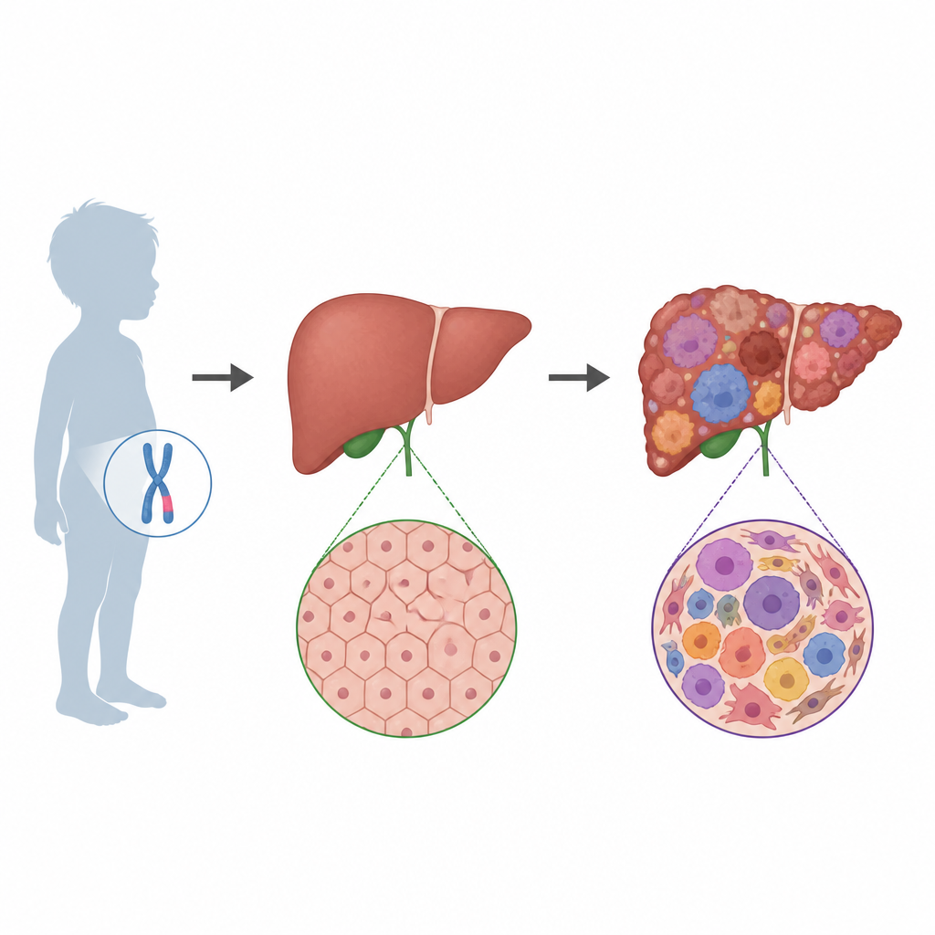
Hepatoblastoma is a rare liver cancer that mostly affects very young children. Some children are born with an overgrowth condition called Beckwith–Wiedemann syndrome, which already puts them at higher risk for tumors. This study uses powerful new tools to zoom in on individual cells from children’s livers and tumors, revealing how normal liver tissue in these high‑risk kids can quietly shift toward cancer. Understanding that hidden journey may eventually help doctors catch tumors earlier and tailor treatments more precisely.
A closer look at a rare overgrowth condition
Beckwith–Wiedemann syndrome, or BWS, is caused by changes in how certain growth genes are controlled on a part of chromosome 11. Children with BWS often grow larger than average and face a higher chance of developing several cancers, including hepatoblastoma. Earlier work showed that even liver tissue that looks normal under the microscope in these children can carry subtle molecular signs of risk. The current study asked what actually happens inside individual liver cells as they move from this risk state to a full‑blown tumor, and how this process compares with liver cancer in children who do not have BWS.
Reading thousands of cells one by one
The researchers collected liver tumors and nearby non‑tumor liver tissue from four children with BWS and three without BWS. Using a “multiomic” approach, they examined both gene activity and the openness of DNA in the nuclei of more than 140,000 cells. This allowed them to sort cells into distinct groups such as normal liver cells, blood vessel cells, immune cells, and several types of tumor cells. They also looked for large‑scale DNA changes that are a hallmark of cancer cells. Together, these layers of information created a detailed map of the cellular “neighborhoods” inside each child’s liver.
Hidden diversity inside tumors
Even when tumors from BWS and non‑BWS patients looked similar under the microscope, their molecular patterns were not the same. Tumors from BWS patients showed stronger signals from growth‑driving pathways, especially the WNT pathway, which is known to push liver cells toward uncontrolled division. These tumors also looked more “embryonic” at the gene level, resembling very early developmental stages rather than more mature liver tissue. In contrast, tumors from children without BWS showed more emphasis on energy use and fat processing. Both groups, however, shared some aggressive features, including activation of certain gene clusters linked with poor outcomes in liver cancers.
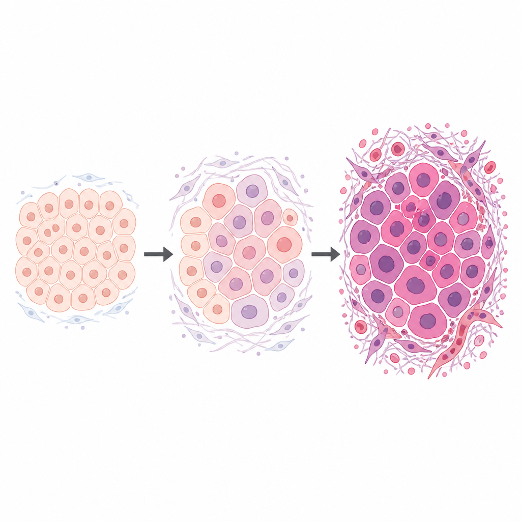
Discovery of transition cells on the road to cancer
By arranging cells along a computed timeline, the team traced a path from healthy‑looking liver cells to fully malignant tumor cells. In BWS samples, this path passed through a distinct “transition” state. Transition cells did not yet have the full genetic chaos of cancer cells, but their gene activity and DNA accessibility showed they were no longer normal either. They began to switch on genes that remodel the tissue scaffold around them and alter how cells contact each other, while true cancer cells went on to strongly activate pathways that drive division and invasion. In non‑BWS samples, intermediate cells were more focused on shifts in metabolism, and the classic cancer signals appeared later.
Signals that may guide future prevention
By combining gene activity, DNA accessibility, and cell‑to‑cell communication patterns, the researchers identified a network of control proteins that seems to steer BWS liver cells from a high‑risk but noncancerous state into these transition cells and then into cancer. Many of the genes in this network influence how cells interact with their surroundings and respond to growth signals. While this work does not yet change clinical care, it points to specific cell types and pathways that might someday be targeted to interrupt tumor development in children with BWS.
What this means for families and clinicians
For families affected by Beckwith–Wiedemann syndrome, this study provides a clearer picture of why liver cancer risk is higher and how tumors may arise from seemingly normal tissue. The findings suggest that a small but important group of transition cells acts as a bridge between healthy liver and cancer, especially in children with BWS. If future research confirms and expands these results, doctors may be able to design tests that detect these early changes or develop treatments that block the key pathways before a tumor takes hold.
Citation: Nirgude, S., Tichy, E.D., Zhang, Y. et al. Beckwith-Wiedemann syndrome multiomic analysis of hepatoblastoma uncovers unique tumour heterogeneity and cellular landscapes, including transition cells leading to tumour formation. BJC Rep 4, 25 (2026). https://doi.org/10.1038/s44276-026-00215-z
Keywords: Beckwith-Wiedemann syndrome, hepatoblastoma, single-cell analysis, WNT signaling, tumor heterogeneity