Clear Sky Science · en
SparStVR - exploring sparse 3D histology data in virtual reality
Seeing Hidden Worlds Inside Tissue
Doctors and researchers often study thin slices of tissue to understand how diseases like cancer grow and spread. But our bodies are three-dimensional, while a microscope slide is flat. This article introduces SparStVR, a virtual reality tool that lets scientists step inside layered tissue images and explore them as lifelike 3D structures, making it easier to spot patterns and problem areas that are hard to see on a regular screen.
From Flat Slices to a 3D Organ
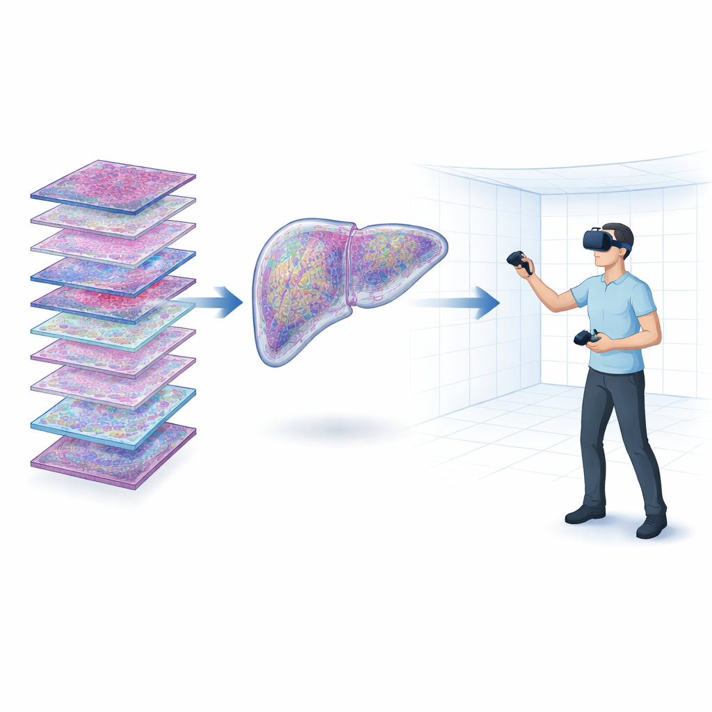
Traditional tissue studies rely on stacks of microscope images taken from many thin cuts of an organ. These stacks can reach gigapixel sizes and are difficult to visualize: most software flattens them into simple projections or lets users rotate a blocky 3D view on a regular monitor. That makes it hard to truly grasp where, for example, a tumor sits inside an organ or how it relates to its surroundings. SparStVR tackles this by turning stacks of images into detailed 3D models that can be walked around and examined inside a virtual environment, giving a natural sense of depth and position.
Walking Around Inside the Data
SparStVR was built with a modern game engine and runs on consumer virtual reality headsets. The user supplies two main ingredients: a stack of color tissue images and corresponding masks that mark structures of interest, such as tumors or glands. The software automatically builds a textured 3D model of the whole sample and the selected structures, placing everything at pixel-accurate locations so that the virtual shapes match the original images closely. Once inside VR, a researcher can grab individual structures, rotate them in mid-air, peel them away from the rest of the organ, and view patches of the original tissue slices exactly where they belong within the 3D form.
Making the Most of Sparse and Mixed Data
In many real studies, only some of the possible tissue sections are stained and scanned, either to save time and cost or to keep material for other tests. This creates “sparse” stacks with gaps between slices, which are especially hard to rebuild as smooth 3D shapes. The authors show that SparStVR still produces convincing organ and tumor models even when only every second, third, or fourth section is available. Smaller details become less precise as more slices are skipped, but the overall organ shape remains realistic. The tool can also combine multiple kinds of images, such as different stains or other measurement maps, allowing a single virtual organ to carry many layers of biological information at once.
Adding Numbers and Signals to the Scene
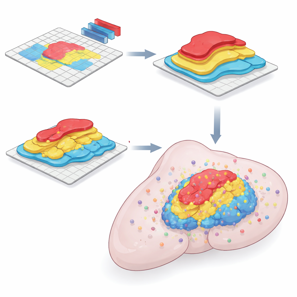
Beyond the visible tissue structure, SparStVR lets users overlay measurements directly into the 3D space. For example, the authors extracted information from the hematoxylin stain channel, which reflects how densely packed cell nuclei are. These values are shown as colored spheres floating at the right locations inside the organ or within specific tumors, turning abstract numbers into a tangible landscape of “hot” and “cold” regions. Additional measurements, such as gene activity or protein levels from modern spatial techniques, can be loaded in the same way, opening possibilities for rich, layered views of disease processes.
Limits, Practicalities, and What Comes Next
While VR opens striking new ways to inspect tissue, it also comes with challenges. Headsets and controllers can cause discomfort for some users, and rendering detailed 3D models from huge image files demands a strong graphics card. The current version of SparStVR runs on Windows PCs that already meet the minimum requirements for VR gaming, and it includes helpful features like easy screenshots and a spectator view for teaching sessions. The authors discuss future directions such as handling many more measurement points, adding built-in automatic segmentation with artificial intelligence, and even simulating disease progression or “digital twins” of organs directly within the virtual space.
Why This Matters for Understanding Disease
By letting researchers virtually step inside a diseased organ, SparStVR turns complex stacks of tissue slices into an intuitive 3D experience. Tumors and other structures can be seen in their true shape and position, and subtle changes in texture or measurement values become easier to notice and interpret. For non-specialists, this means that the path from a flat microscope slide to an understanding of how a disease occupies and reshapes an organ becomes more direct and concrete. As VR and AI tools advance, approaches like SparStVR could help transform how scientists, students, and clinicians explore the hidden architecture of the body.
Citation: Liimatainen, K., Latonen, L. & Ruusuvuori, P. SparStVR - exploring sparse 3D histology data in virtual reality. Commun Eng 5, 79 (2026). https://doi.org/10.1038/s44172-026-00634-3
Keywords: virtual reality, 3D histology, cancer imaging, tissue visualization, digital pathology