Clear Sky Science · en
Comprehensive evaluation of cross cancer generalization in histopathology segmentation models across 21 tumor types
Smarter helpers for cancer diagnosis
Doctors who study tissue under the microscope increasingly rely on computer programs to spot cancer and measure it precisely. But building a separate tool for every single cancer type takes months of expert work. This study asks a simple question with big practical impact: can a high-quality tool trained for one cancer be safely reused for many others, saving time and speeding up access to digital helpers in real clinics? 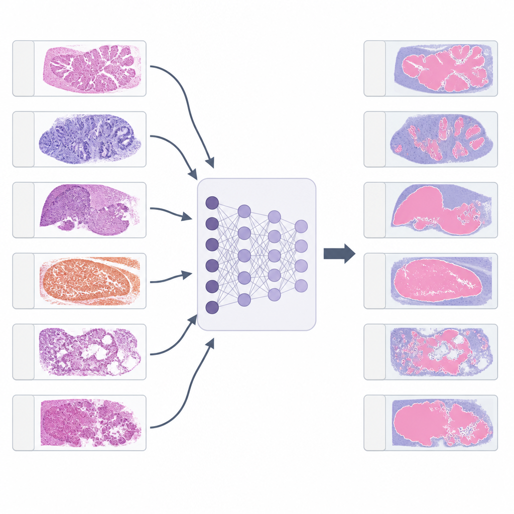
Why marking cancer on slides matters
When a pathologist looks at a tissue slide, they do not just ask if cancer is present. They carefully trace the borders between tumor and healthy tissue, judge how the cells are arranged, and measure features such as scar-like tissue, dead areas, and immune cells. Modern artificial intelligence can mimic this process by coloring every pixel of a slide according to what it contains. This pixel-level mapping supports research on how tumors grow, helps quantify subtle patterns that humans struggle to measure, and can feed into new tests that link tissue structure to genetic or drug response data.
The challenge of one-model-per-cancer
Creating such detailed tools is slow and demanding. Experts must draw precise outlines of tumor and surrounding tissue on many large images, a task that can take several hours per case. For each new cancer type, a fresh model is usually trained and then tested for safety and accuracy, often over years and across many hospitals. With dozens of common tumor types, this “one model per cancer” approach becomes a serious bottleneck. It limits how quickly artificial intelligence can be brought into routine care and makes it harder to build widely usable systems.
Testing whether models can cross cancer borders
The researchers took five existing tissue segmentation models originally trained for breast, colon, lung, kidney, and prostate cancer. They applied them to more than 7,700 digital slides from 21 different tumor types collected by The Cancer Genome Atlas project. For each slide, a pathologist selected one large region rich in tumor and, when possible, one region of benign tissue. The models colored every pixel in these regions according to tissue type. Pathologists then scored how well each model separated tumor from its supporting tissue on a simple 0 to 10 scale, where higher scores meant more accurate outlines and fewer missed structures. 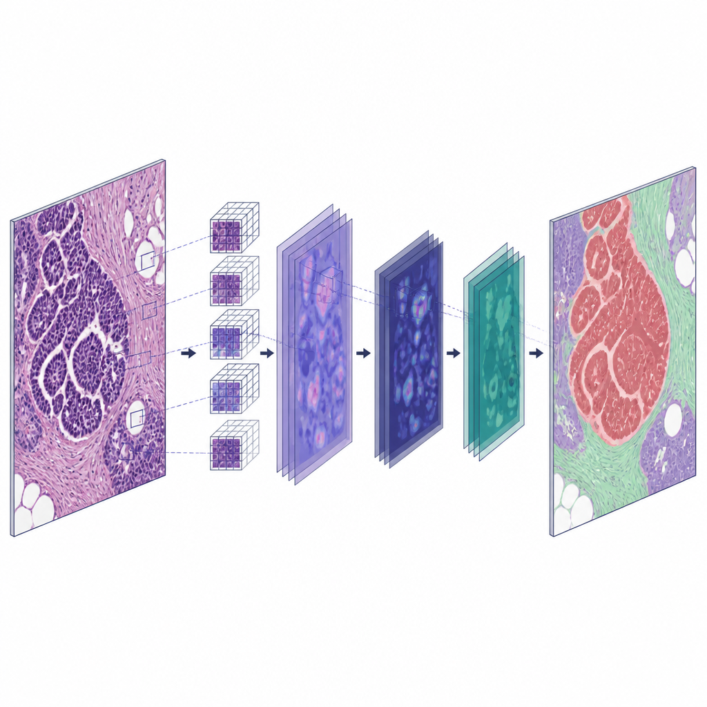
Which models traveled best across cancers
The lung model turned out to be the most widely useful. On average it scored about 8 out of 10 across all tumor types, and it reached “excellent” quality in more than half of the non-lung cancers tested, including ovarian tumors, bile duct tumors, thyroid cancer, and several cancers built from flat squamous cells. In many of these settings, its performance matched that seen in its original lung slides. Breast and colon models also worked well for many other cancers, though not as broadly as the lung model. Kidney and prostate models, trained on more narrowly shaped tumors, generalized less reliably, especially for cancers whose cells look very different under the microscope.
Patterns that explain good generalization
The study revealed that success or failure largely followed how similar the cell shapes and growth patterns were between tumors. For example, the lung model, which had learned both gland-forming and squamous growth in lung cancers, worked well on squamous cancers of the head and neck, cervix, and esophagus. The breast model, trained to detect scattered single cells in a subtype called invasive lobular carcinoma, proved effective for diffuse stomach cancers made of similar loose, small cell clusters. By contrast, tumors with unusual appearances, such as many kidney cancers and melanomas, were harder for all models. Even so, the tools often produced useful outlines in benign regions, such as normal glands or early pre-cancer changes, which could be repurposed in future training.
Faster paths to new clinical tools
Because several models already work well on cancers they were never shown during training, they can serve as “starter tools” rather than forcing experts to label everything from scratch. The authors outline practical routes: directly using a strong model without retraining for some studies, using its output as a first draft that pathologists quickly correct, or converting older, coarse datasets into precise pixel maps by combining patch labels with these segmentations. In favorable cases, this can shrink the time needed to build a new model from more than a year to under a week. For patients, this could mean that accurate, explainable artificial intelligence supports diagnosis and biomarker discovery across many cancers sooner and with less duplicated effort.
What this means for future cancer care
For a lay reader, the key message is that the same smart computer program used to trace lung tumors can often be reused to outline many other cancers without starting from zero. This reuse trims the slowest part of model building, which is careful manual drawing by experts, and helps researchers focus on testing and improving tools instead of redrawing the same patterns over and over. If developed further into true “all-cancer” systems, such segmentation models could become a standard layer in digital pathology, providing consistent maps of tumor and surrounding tissue that feed into better studies, clearer reports, and, ultimately, more informed clinical decisions.
Citation: Bedau, T., Harder, C., Al-Shughri, A. et al. Comprehensive evaluation of cross cancer generalization in histopathology segmentation models across 21 tumor types. Commun Med 6, 302 (2026). https://doi.org/10.1038/s43856-026-01601-x
Keywords: computational pathology, cancer segmentation, deep learning, digital histology, pan-cancer models