Clear Sky Science · en
Physics-based machine learning toolbox for probing concentration under diffusive regime in microfluidics devices
Watching tiny chemistry in action
When liquids weave through the microscopic pores of rocks, they can carry pollutants, store carbon dioxide, or lock away nuclear waste. Scientists use microfluidic chips—tiny, transparent devices that mimic porous rocks—to watch these processes unfold. But while cameras can capture beautiful movies of crystals growing and pores clogging, they cannot directly show how dissolved chemicals move and build up inside the channels. This article introduces a new computer-based toolbox that turns images from these chips into fast, quantitative maps of chemical concentrations, helping researchers see the invisible and react to experiments in real time.
From pictures to hidden concentration maps
The authors tackle a practical bottleneck in modern microfluidics: how to measure changing concentrations of dissolved substances across a complex chip without adding slow or invasive sensors. Traditionally, scientists run detailed numerical simulations, which act like virtual probes. These simulations are accurate but extremely time-consuming and require painstaking manual preparation of each image. The new toolbox replaces most of this heavy lifting with a physics-based machine learning approach. It takes in regular optical microscopy images and physical conditions and rapidly predicts how different dissolved species are distributed in space and time, as well as how quickly they diffuse and where solid crystals are likely to appear.
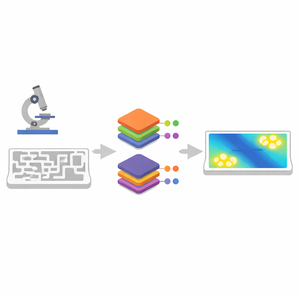
Three smart steps instead of one big guess
Rather than relying on a single opaque neural network, the toolbox mimics the usual modeling workflow in three transparent steps. First, a U-Net model—a type of image-segmentation network—converts raw color images from the microscope into simple black-and-white maps that distinguish solid grains from pore space. This removes the need for laborious manual tracing of each grain boundary. Second, a convolutional autoencoder compresses these binary images into a compact set of numbers that still capture the essential geometry. It also generates many additional realistic pore patterns, expanding the training set without more experiments or manual work.
Embedding physics for speed and trust
The final step uses a technique called a non-intrusive reduced basis method. Instead of treating the system as a black box, this method starts from full physics simulations based on the Lattice–Boltzmann approach—well-established equations for how particles diffuse through complex structures. From these simulations, the method extracts a small set of basis patterns that describe how concentration fields typically behave. A lightweight neural network then learns only how the compressed geometry, time, and physical settings combine to weight these patterns. This design keeps the underlying physics intact while slashing computation time: once trained, the toolbox can replace simulations that would take many hours on a supercomputer with predictions computed in a fraction of a second on a laptop.
Testing on evolving rock-like and uniform chips
To demonstrate the toolbox, the team studied two microfluidic experiments where solutions containing strontium and sulfate mix and form crystals of celestine (SrSO₄). One chip imitated a natural rock with irregular grains; the other used a regular pattern of circular grains. In both cases, repeated optical images captured how pores slowly clogged as crystals grew. Using only a handful of manually segmented images for training, the U-Net and autoencoder achieved segmentation accuracies above 97–99%. The reduced-basis models then reproduced full simulation results for both tracer diffusion and four different ionic species with tiny errors, while being more than one hundred thousand to a million times faster than the original simulations.
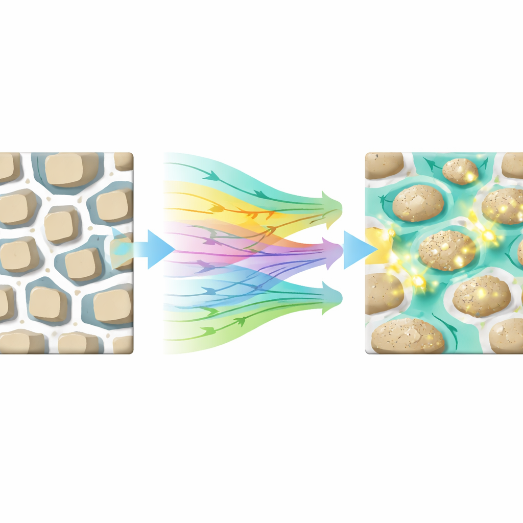
Pointing to where crystals will grow next
Armed with fast concentration predictions, the toolbox can calculate a saturation index—a measure of whether local conditions favor crystal growth. The authors used this to forecast, ten minutes in advance, where new celestine crystals would appear or existing ones would grow in both chips. Regions flagged as supersaturated by the model closely matched the locations where bright new crystals later emerged in the microscope images. At the same time, the toolbox estimated how the overall ease of diffusion through the chip changed as pores clogged, providing a bridge between microscopic structure and macroscopic transport properties.
Why this matters for future experiments
In plain terms, this work turns microfluidic chips into smarter, partly digital experiments. By combining machine learning with physical insight, the toolbox gives researchers a way to "see" concentration fields and likely precipitation spots almost as quickly as images are recorded, without giving up interpretability. This opens the door to true digital twins of microfluidic systems: virtual copies that run alongside real experiments and suggest when to change conditions, where to focus advanced imaging beams, and how evolving pore structures affect flow and reaction. While current results focus on diffusion-driven reactions, the same framework could eventually help design and control a wide range of lab-on-a-chip processes, from carbon storage to groundwater cleanup.
Citation: Santoso, R., Yang, Y., Lönartz, M. et al. Physics-based machine learning toolbox for probing concentration under diffusive regime in microfluidics devices. Commun Phys 9, 106 (2026). https://doi.org/10.1038/s42005-026-02590-y
Keywords: microfluidics, physics-based machine learning, porous media, crystal precipitation, digital twin