Clear Sky Science · en
De-aberration for noninvasive transcranial photoacoustic computed tomography through an adult human skull
Seeing the Brain Without Opening the Skull
Doctors and scientists dream of watching the brain at work in real time, without bulky MRI scanners or opening the skull. Photoacoustic computed tomography is a promising technology that uses flashes of light and sensitive microphones to map blood in the brain. But the adult human skull badly distorts these sound waves, blurring the images and hiding important details. This study shows, for the first time in realistic experiments with adult human skulls, that those distortions can be largely undone—bringing us closer to a practical, noninvasive brain-imaging tool that could someday sit at the bedside.
How Light and Sound Can Map the Brain
Photoacoustic imaging works in a simple but powerful way. Short laser pulses are aimed at tissue, where blood strongly absorbs the light and heats up by a tiny amount. That rapid heating causes the blood to emit ultrasound waves, which travel outward and are picked up by an array of detectors. Because different forms of hemoglobin absorb light differently, this method can track blood oxygen and blood flow—key indicators of brain activity—more directly than conventional MRI, and with a smaller, quieter, and cheaper setup. In soft tissues like breast or limb, the sound waves travel smoothly, and standard mathematical methods can accurately rebuild the picture. The skull, however, is a different story.
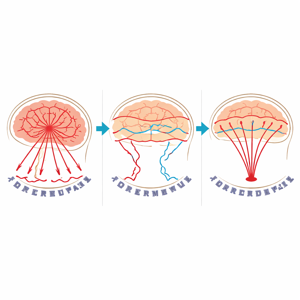
Why the Skull Blurs the Picture
The human skull has very different mechanical properties from the brain and surrounding soft tissues. It is stiffer, denser, and supports not only the usual squeeze-like compression waves but also sideways shear waves. When the photoacoustic waves from the brain hit the skull, part of the energy is reflected, part is converted between these two wave types, and all of it is slowed and bent in complicated ways. On top of that, the skull weakens higher-pitched sound more than lower-pitched sound. Conventional reconstruction methods treat the head as if it were filled with one uniform material, so they fail dramatically when the waves have been twisted and delayed by bone. Images of brain-mimicking targets behind a real skull turn into unrecognizable smears.
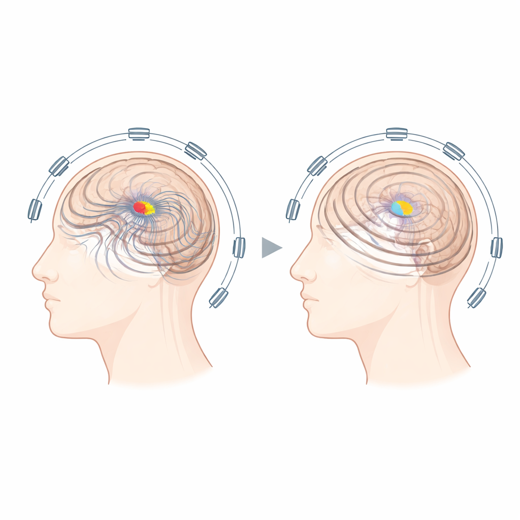
A New Way to Undo the Distortion
The authors tackled this long-standing problem by explicitly modeling the skull as an elastic solid instead of ignoring it. They first obtained the 3D shape and placement of ex-vivo adult human skulls using standard medical scans such as CT or MRI, then assumed that the bone inside that outline could be treated as a single, uniform material with realistic sound speeds. Using a powerful full-wave simulation, they calculated how sound would travel from many possible source points inside the skull to the detector array. An iterative computer algorithm then searched for the pattern of initial pressure inside the skull that best matched the measured signals, while respecting basic assumptions such as non-negative signal strength and smoothness.
Clearer Images Across Setups and Skulls
To test the method, the team placed thin, blood-filled tubes, black wires, and 3D-printed shapes just inside the skulls to mimic brain vessels. They compared images taken without the skull, with the skull but reconstructed in the usual way, and with the skull but reconstructed using their new model. Standard reconstructions with the skull in place were so distorted that the patterns were barely recognizable. In contrast, the new approach recovered the fine branching structures and positions of the targets with striking fidelity, whether the light came from inside the skull cavity or from outside, as would be required clinically. The improvement held across different depths, different target shapes, and even across two separate skulls obtained from different donors. The researchers also deliberately introduced errors into the assumed skull position, orientation, and sound speeds, and found that while ignoring shear waves crippled performance, modest inaccuracies in the other parameters still yielded useful images.
What This Means for Future Brain Scans
This work shows that skull-induced blurring in photoacoustic brain imaging is not an insurmountable barrier. With only the skull’s shape, position, and orientation—information that hospitals can already obtain from CT or specialized MRI scans—the new method can refocus scrambled sound waves and recover sharp images behind adult human skulls, at least in controlled experiments. Although further challenges remain, such as handling signals from scalp structures and fully accounting for the skull’s internal variations, the study demonstrates a realistic path toward a portable, radiation-free imaging tool that could complement MRI for monitoring strokes, injuries, and other brain conditions at the bedside.
Citation: Aborahama, Y., Sastry, K., Cui, M. et al. De-aberration for noninvasive transcranial photoacoustic computed tomography through an adult human skull. Commun Phys 9, 116 (2026). https://doi.org/10.1038/s42005-026-02545-3
Keywords: photoacoustic brain imaging, transcranial imaging, skull aberration correction, functional neuroimaging, ultrasound and light