Clear Sky Science · en
Single cell and spatial sequencing analysis of cancer associated fibroblasts in the brain metastasis tumor microenvironment
Why brain tumors and their neighbors matter
When cancers from the lung or breast spread to the brain, they are very hard to treat and often deadly. These tumors do not grow alone: they are surrounded by normal brain cells, blood vessels, and a little-known group of support cells called fibroblasts. This study looks closely at those fibroblasts in human brain metastases to see how they are organized, how they talk to other cells, and whether some might actually help keep tumor growth in check.
The crowded neighborhood inside brain tumors
The researchers used advanced single cell and spatial sequencing on nine brain metastasis samples taken from patients with lung or breast cancer. These tools allowed them to profile tens of thousands of individual cells and map where they sit in the tissue. They found that each tumor contains a mix of cancer cells and many types of noncancer cells, including immune cells, blood vessel cells, nerve cells, and cancer associated fibroblasts. Fibroblasts turned out to be among the most common noncancer cells, suggesting they play an important role in how these tumors develop and respond to therapy.
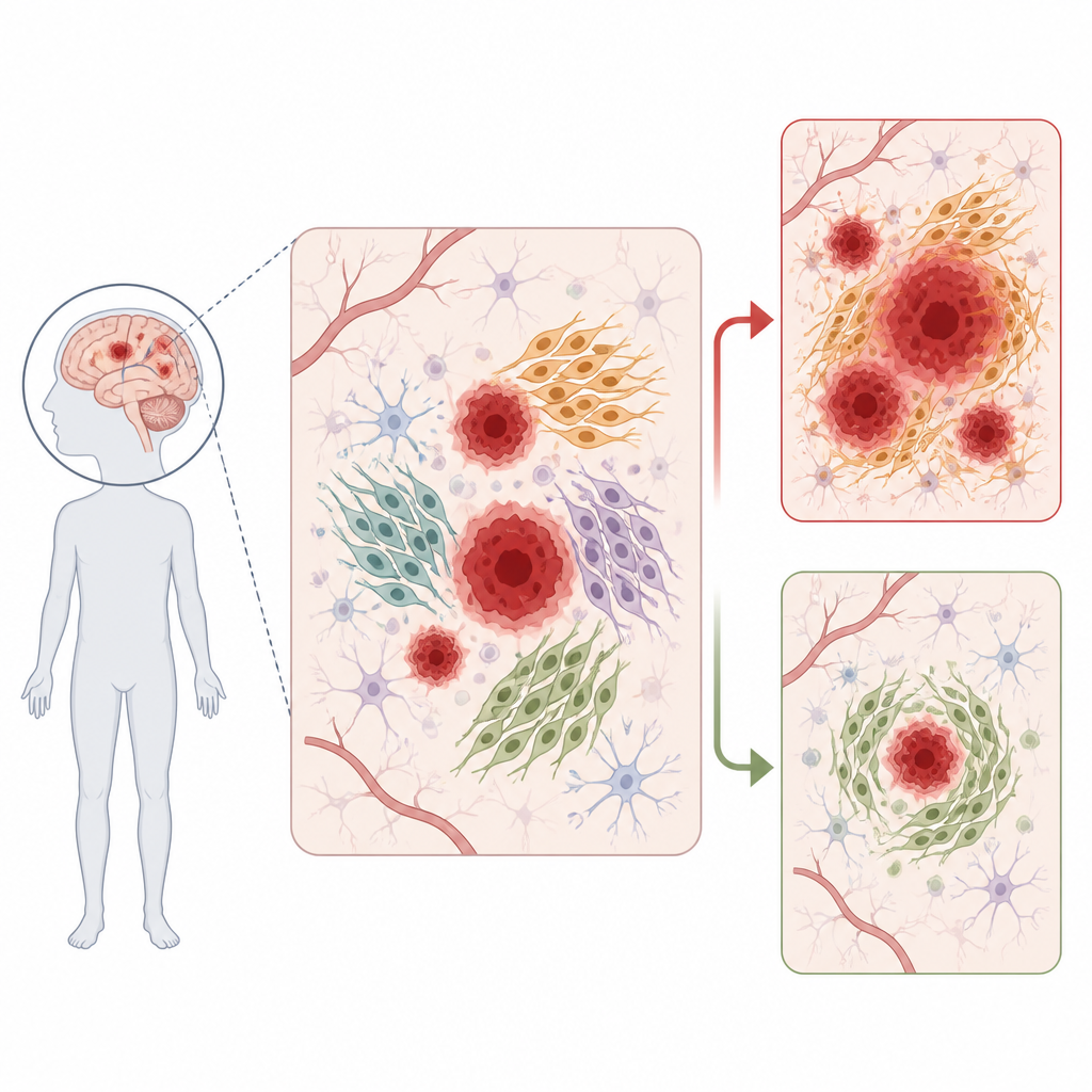
Four kinds of helper cells around the tumor
By focusing on the fibroblasts, the team discovered that they are not all the same. Instead, they fall into four clear groups based on which genes are active. One group builds and organizes the tissue scaffold that surrounds cells, another shows strong links to immune functions, a third has features related to muscle like contraction, and a fourth carries markers normally seen in nerve cells and appears specific to the brain. These four groups are found in different proportions in each patient and tend to favor certain brain regions, hinting that local brain structure helps shape which fibroblasts appear.
Cellular conversations and tissue layout
The scientists then asked how these fibroblast groups communicate with other cells. Using a database of known signaling pairs, they showed that fibroblasts exchange many signals with cancer cells, immune cells, blood vessels, and neighboring brain cells. Different fibroblast groups use different signal combinations, consistent with specialized roles such as promoting new blood vessel growth, modulating immune activity, or interacting with neurons and supporting cells of the brain. Spatial gene maps of four tumors confirmed that fibroblasts occupy stromal bands next to, but largely separate from, dense cancer cell zones, while immune cells move more freely into tumor rich regions.
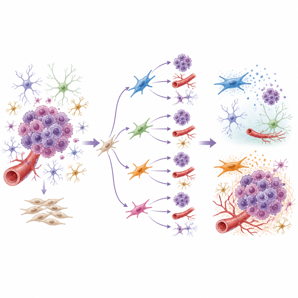
Hidden layers within each fibroblast group
Within each of the four main fibroblast types, the study found smaller subclusters with distinct activity patterns. Some subclusters showed hallmarks of supporting tumor growth, such as promoting inflammation, stiffening the surrounding tissue, or fueling blood vessel changes. Others looked more like normal support cells. One particular subcluster, within the tissue scaffold building group, stood out because it expressed a protein called ISLR that has been linked in other cancers to restraining tumor growth and limiting tissue scarring. Trajectory analysis suggested that this ISLR rich state may arise from more normal fibroblast like precursors.
Fibroblasts that can slow tumor cells
To test whether ISLR rich fibroblasts in brain metastases can actually affect tumor behavior, the authors used patient derived fibroblast cell lines grown in the lab. They compared cell lines with high or low ISLR levels and collected the substances these fibroblasts release into their surroundings. When they exposed brain metastasis tumor cells to this conditioned fluid, the secretions from high ISLR fibroblasts reduced tumor cell viability over several days, whereas one low ISLR line even boosted tumor cell growth. These results support the idea that at least some fibroblasts in brain metastases can act as natural brakes on tumor expansion.
What this means for future treatments
This work paints a detailed picture of fibroblasts in brain metastases, revealing four major types and many subtypes that differ in where they sit, how they signal, and whether they are likely to help or hinder tumor progression. For patients, the key message is that not all stromal cells around a tumor are bad. Some, such as the ISLR rich fibroblasts, may actually fight against the cancer. Therapies that broadly wipe out fibroblasts could therefore remove helpful cells along with harmful ones. A more refined approach that targets only tumor supporting fibroblast subtypes, while preserving or even boosting tumor inhibiting ones, may offer a smarter way to reshape the tumor environment and improve outcomes for people with brain metastases.
Citation: Simon, T., Buckley, D.N., Yang, Z. et al. Single cell and spatial sequencing analysis of cancer associated fibroblasts in the brain metastasis tumor microenvironment. Commun Biol 9, 714 (2026). https://doi.org/10.1038/s42003-026-09915-1
Keywords: brain metastasis, tumor microenvironment, cancer associated fibroblasts, single cell sequencing, ISLR fibroblasts