Clear Sky Science · en
Interplay between DYRK1A dosage and aneuploidy-induced neuropathology in Down syndrome
Why this research matters for families and caregivers
People with Down syndrome often face learning difficulties and a higher risk of early memory loss, but the reasons why brain cells are so vulnerable have remained unclear. This study uses human stem cell models to uncover how two hidden problems inside the brain’s cells team up to damage neurons, and it suggests a combined treatment strategy that might one day help protect thinking and memory in Down syndrome. 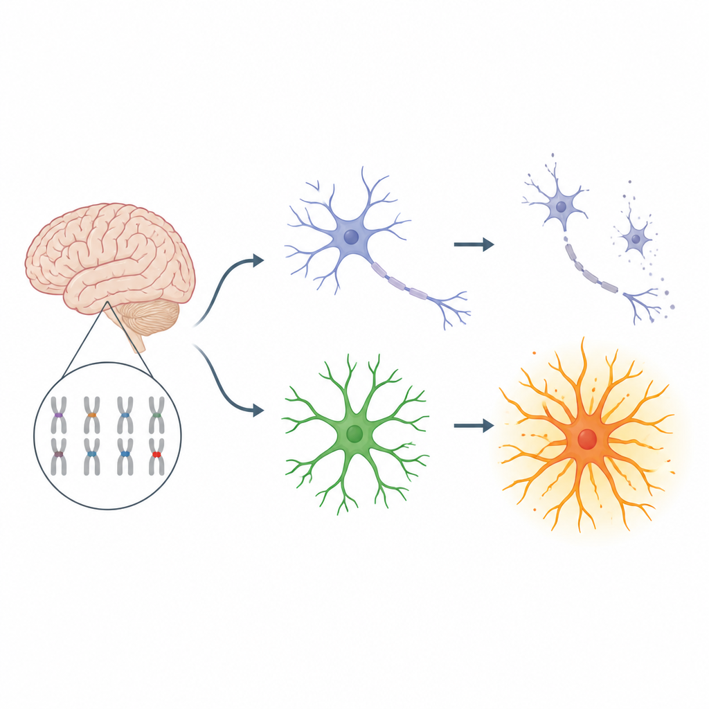
Two kinds of brain cells caught in a harmful partnership
The brain relies on close teamwork between neurons, which send signals, and astrocytes, star-shaped support cells that guide and protect neurons. In Down syndrome, an extra copy of chromosome 21 shifts this balance: there are fewer neurons and roughly twice as many astrocytes as usual, and the astrocytes tend to stay in a chronically “switched on” state. The researchers asked how stress inside trisomy brain cells and extra copies of certain genes on chromosome 21 might combine within this neuron–astrocyte partnership to drive long-term damage.
Stressed neurons burdened by clumped proteins
Using induced pluripotent stem cells from people with trisomy 21, 18, and 13, the team grew neurons in the lab and compared them with matched cells that had been corrected back to the usual chromosome number. Across all three trisomies, neurons showed strong signs of internal stress: proteins misfolded and clumped into aggregates, and more cells underwent programmed cell death. A key protein involved in Alzheimer’s disease, tau, was found in an overly phosphorylated, aggregation-prone form, and this altered tau leaked into the fluid around the neurons. Treating the neurons with a chemical “chaperone” called 4‑phenylbutyrate reduced these protein clumps and lowered the amount of stressed tau released, showing that the chromosomal imbalance itself can trigger a common type of toxic protein buildup. 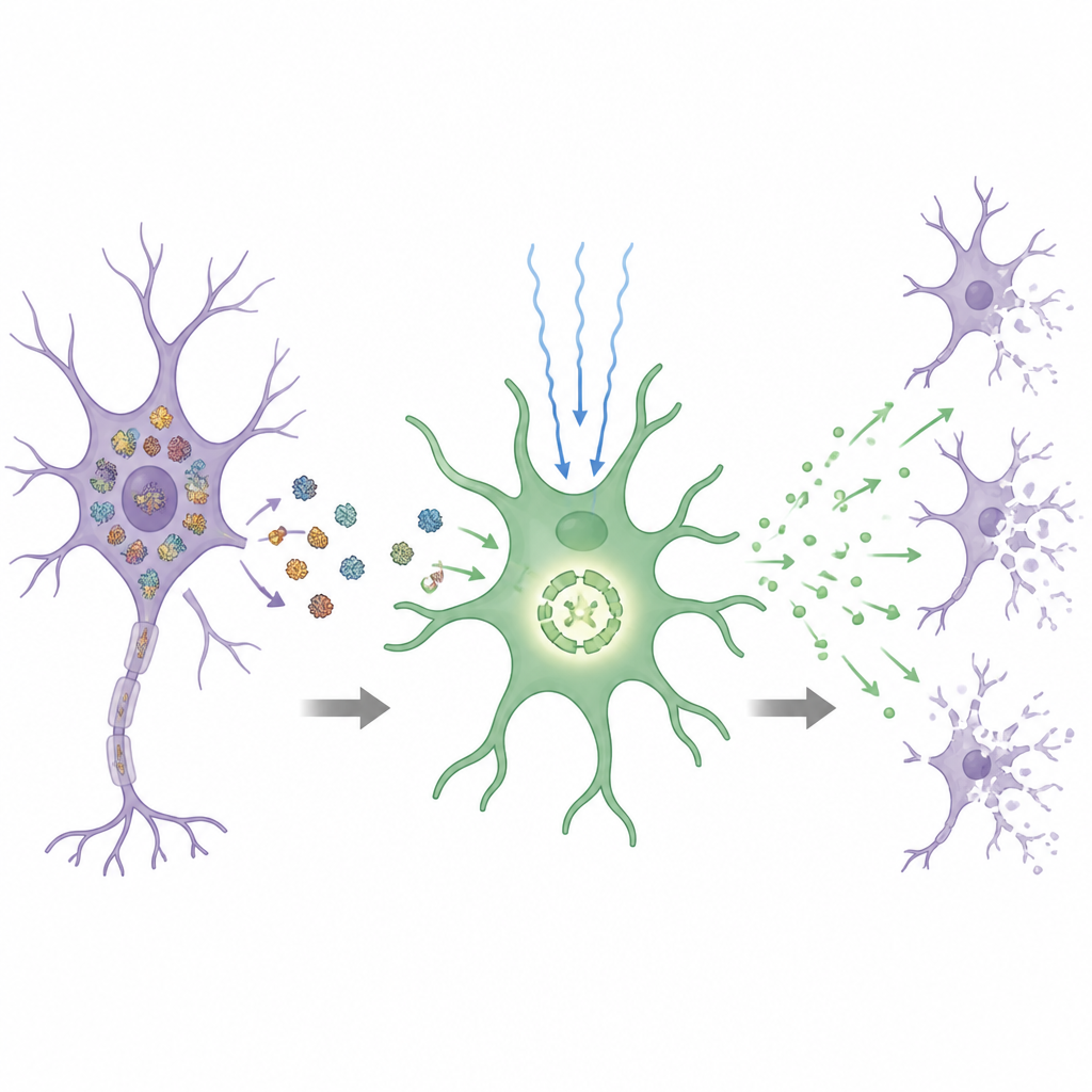
Astrocytes in Down syndrome become natural fire starters
The story was different for astrocytes. Only trisomy 21 astrocytes showed unusually rapid growth and a strong inflammatory profile, pointing to a specific effect of the extra chromosome 21 rather than general chromosomal stress. These astrocytes had an internally activated danger sensor called the NLRP3 inflammasome, which normally helps control the release of inflammatory substances. Even without standard priming signals, trisomy 21 astrocytes produced high levels of inflammatory molecules such as interleukin‑1β, and this response could be dialed down with drugs that block NLRP3. When neurons were grown together with trisomy 21 astrocytes, protein clumps and cell death in neurons rose sharply, showing that these reactive support cells can turn from protectors into drivers of damage.
A vicious loop powered by a single extra gene
The researchers next asked which chromosome 21 gene pushes astrocytes toward this inflammatory state. By studying a cell line missing only a small critical region of chromosome 21, and then selectively silencing individual genes, they pinpointed DYRK1A, a gene already known for its role in brain development. Extra DYRK1A boosted key signaling pathways inside astrocytes that feed into NLRP3, while reducing DYRK1A activity with genetic correction or drugs calmed expression of NLRP3 and its inflammatory products. At the same time, medium collected from stressed trisomic neurons further heightened the inflammatory output of trisomy 21 astrocytes, and this effect faded when neuronal protein stress was relieved by 4‑phenylbutyrate. Together, these findings reveal a feed‑forward loop: chromosomal stress causes neurons to shed toxic protein forms, which activate DYRK1A‑sensitized astrocytes, which in turn release signals that make neurons even more likely to die.
Combining two levers to better protect neurons
Because the damage loop has two main drivers, the team tested a two-pronged strategy. Correcting DYRK1A copy number only in astrocytes reduced inflammatory signaling and lowered neuronal stress, but neurons with trisomy 21 still died more often than corrected controls. When the researchers combined astrocyte DYRK1A correction with 4‑phenylbutyrate treatment to ease protein stress in neurons, both protein aggregation and neuron death were brought close to normal levels. For a lay audience, the take-home message is that in Down syndrome, neuron loss may stem from a partnership between stressed neurons and overactive support cells, and that calming both sides at once—by tuning down DYRK1A in astrocytes and easing protein stress in neurons—could form the basis of future therapies aimed at preserving brain health.
Citation: Nambara, T., Lee, J.Y., Minami, M. et al. Interplay between DYRK1A dosage and aneuploidy-induced neuropathology in Down syndrome. Commun Biol 9, 660 (2026). https://doi.org/10.1038/s42003-026-09902-6
Keywords: Down syndrome, neurons, astrocytes, brain inflammation, protein aggregates