Clear Sky Science · en
Parallel CRISPR screens reveal pathways controlling the cell surface levels of the attractant receptor FPR1
How Immune Cells Tune Their Sensitivity
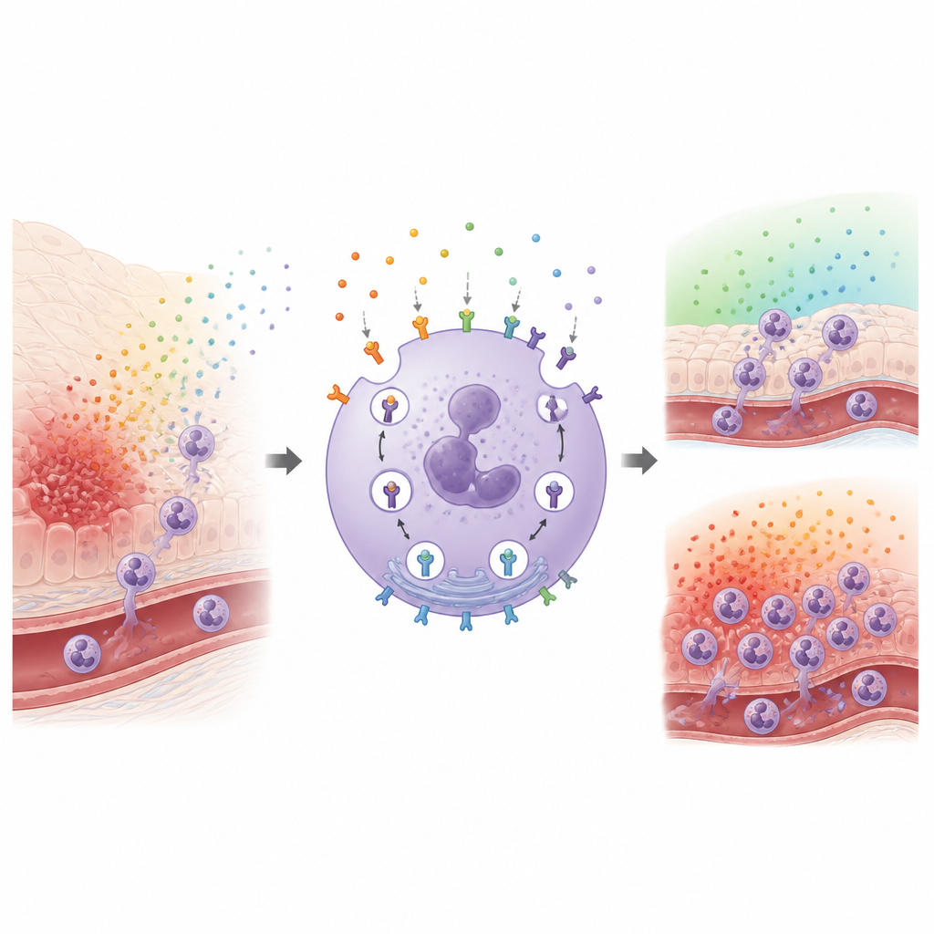
Our immune system relies on front line cells called neutrophils that race toward sites of infection, guided by chemical trails. To avoid either sluggish defense or runaway inflammation, these cells must carefully control how many "smell receptors" sit on their surface. This study asks a simple but important question: how do neutrophils decide when to pull these receptors inside the cell and when to send more back out, and what happens inside the cell during this constant traffic?
The Watchful Gatekeeper on Neutrophils
A key player in this story is a receptor called FPR1, which sits on the surface of neutrophils and senses tiny fragments released by bacteria and damaged tissue. When FPR1 detects these alarm signals, it helps neutrophils migrate toward danger and switch on weapons that can kill microbes but also damage healthy tissue. The number of FPR1 receptors on the surface strongly influences how sensitive a neutrophil is. After activation, many receptors are pulled into the cell, lowering sensitivity, while under other conditions more receptors are moved to the surface, priming the cell to respond. Yet the detailed machinery that adds and removes FPR1 from the cell surface has been surprisingly murky.
Measuring Receptor Traffic in Living Cells
The researchers first refined a way to watch FPR1 move in large numbers of cells at once. They used a neutrophil-like cell line and tagged surface FPR1 with fluorescent antibodies, then stimulated the receptors with a bacterial mimic. By tracking how quickly the surface signal dropped, they could infer how rapidly receptors were being internalized. They confirmed these measurements with microscopy that showed fluorescent attractant accumulating inside cells as surface staining faded. Within minutes, most FPR1 moved off the membrane, revealing that this receptor is cleared quickly and efficiently, with some of it later recycled back to the surface.
Uncovering Parallel Routes into the Cell
Next, the team examined known regulatory proteins. They showed that several receptor-targeting enzymes, called GRKs, work together to tag FPR1 so it can be pulled inward, and that two adapter proteins known as beta arrestins aid this process. However, even when both beta arrestins were removed, FPR1 still internalized partway, implying that at least one additional route into the cell operates in parallel. To systematically search for all the players, the researchers turned to genome-wide CRISPR screening, disrupting almost every gene in the genome in huge pools of cells. They ran two linked screens: one captured genes that affect FPR1 levels on resting cells, and the other captured genes that change FPR1 levels after stimulation, allowing them to distinguish problems in making, moving, removing, or recycling the receptor.
Hidden Machinery of Receptor Recycling
By comparing these screens, the authors mapped a network of pathways that govern FPR1 traffic. They highlighted large protein assemblies that help fold FPR1, carry it through the cell’s internal shipping routes, and sort it in endosomes, the way-stations for incoming cargo. Some complexes, such as retromer, retriever, and CCC, appeared to influence both the baseline amount of FPR1 on the surface and what happens to it after it is internalized. The study also pointed to machinery that moves storage granules to the membrane, enabling bursts of FPR1 delivery when cells encounter attractant. This integrated view shows that receptor levels are not controlled by a single switch but by a layered system of production, routing, and recycling.
New Molecules Steering Receptor Uptake
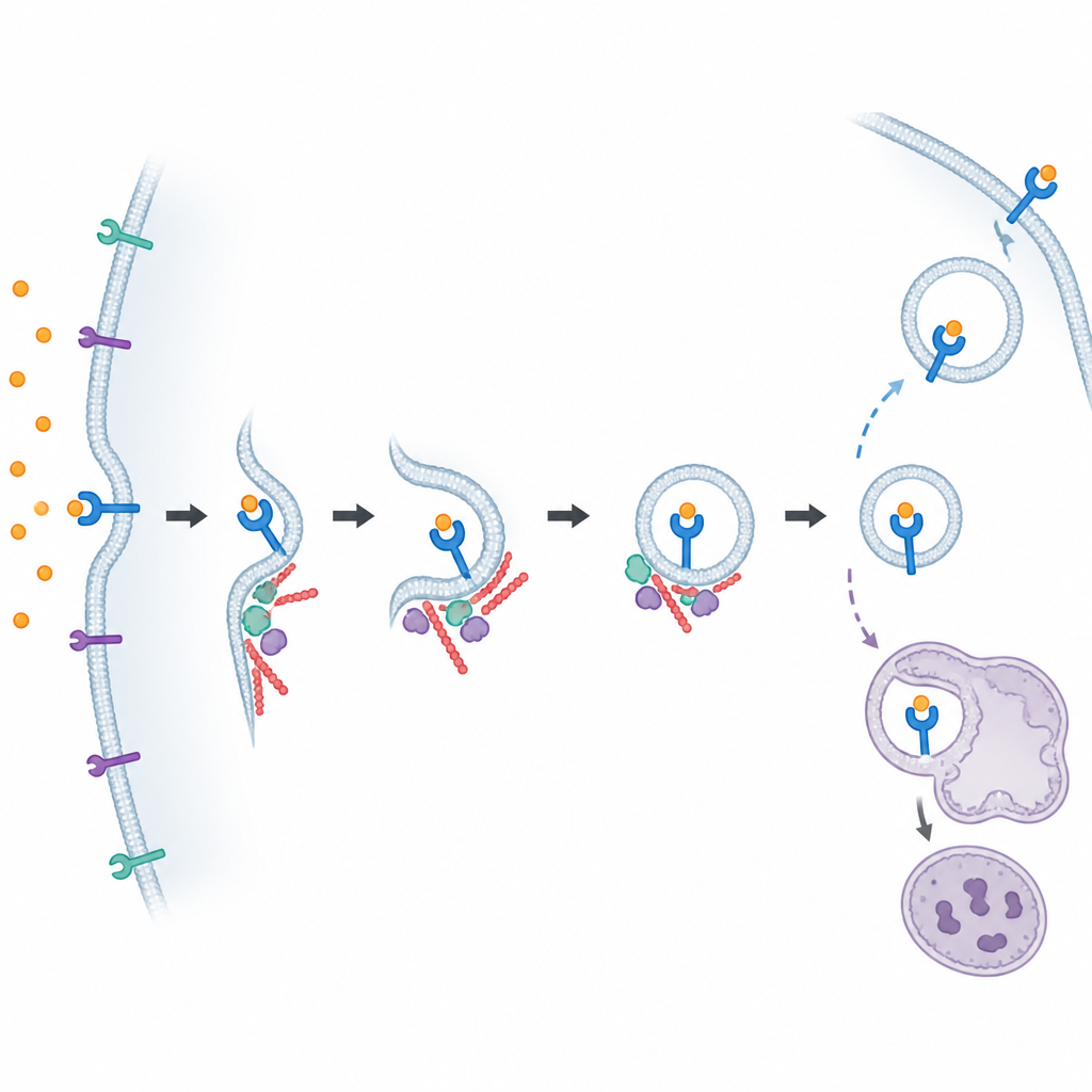
Among many hits, two previously underappreciated proteins stood out as especially important for pulling FPR1 off the surface. One, mDia1, helps build straight actin filaments, part of the cell’s internal scaffolding. The other, ARF6, is a small molecular switch that influences how the membrane bends and how vesicles form. When the team chemically inhibited mDia1 or ARF6, or genetically removed ARF6, neutrophil-like cells and primary human neutrophils failed to internalize FPR1 properly. Further experiments suggested that ARF6 participates particularly in the arm of the pathway that does not rely on beta arrestins, reinforcing the idea that FPR1 uses more than one internalization route.
Why This Cellular Traffic Map Matters
For non-specialists, the takeaway is that neutrophils adjust their "volume knob" for danger signals by tightly managing how many FPR1 receptors are exposed on their surface, using several overlapping pathways. This study provides a map of the proteins and complexes that move FPR1 on and off the membrane, and reveals that actin-building factors like mDia1 and the membrane regulator ARF6 are key parts of this system. Understanding these routes could eventually help scientists design treatments that temper harmful inflammation or harness rapid receptor internalization for targeted drug delivery, by gently nudging the cell’s own traffic control rather than shutting signals off outright.
Citation: Akdoğan, E., Lundgren, S.M., Kamber, R.A. et al. Parallel CRISPR screens reveal pathways controlling the cell surface levels of the attractant receptor FPR1. Commun Biol 9, 668 (2026). https://doi.org/10.1038/s42003-026-09878-3
Keywords: neutrophils, FPR1 receptor, CRISPR screen, receptor endocytosis, immune signaling