Clear Sky Science · en
β-tubulin phosphorylation by Chk1 is required for normal spindle formation during cell division
How Cells Keep Their Genetic Cargo in Order
Every time a cell divides, it faces a high‑stakes engineering challenge: it must build a tiny machine that pulls copied chromosomes apart cleanly so each daughter cell gets the right genetic cargo. When this machine, called the spindle, malfunctions, the result can be birth defects, developmental disorders, or cancer. This study uncovers an unexpected way in which a well‑known DNA damage protein, Chk1, moonlights as a construction supervisor for the spindle itself.
The Cell’s Rope-and-Pulley System
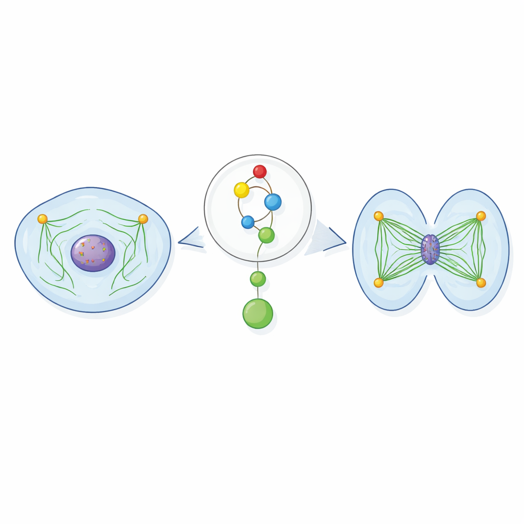
During cell division, long protein fibers called microtubules form the spindle, a bipolar structure that grabs chromosomes and hauls them to opposite sides of the cell. These fibers grow out from organizing centers known as centrosomes and hook onto specific chromosome sites. For this to work, the spindle needs enough microtubules, arranged in the right geometry, to capture and move every chromosome. If the spindle is too sparse or unstable, chromosomes can lag behind, attach incorrectly, or end up in the wrong daughter cell, a state known as aneuploidy that is strongly linked to cancer.
A DNA Guardian with a Second Job
Chk1 is best known as part of the cell’s damage‑response system: when DNA is harmed or replication stalls, Chk1 pauses the cell cycle so repairs can be made. The authors of this paper asked whether Chk1 might also act during normal, undisturbed cell division. By reducing Chk1 levels or blocking its activity in several vertebrate cell lines, they found that spindles still formed but were noticeably thinner, with fewer microtubules radiating from the centrosomes. These defects appeared even when DNA was intact and when other known Chk1‑controlled steps, such as entry into mitosis or activation of another mitotic regulator, Aurora B, were held constant. This showed that Chk1 has a distinct, direct role in building a robust spindle.
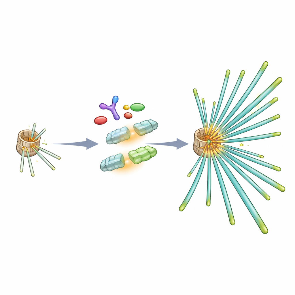
Switching On Tubulin to Build a Stronger Spindle
To understand how Chk1 reinforces the spindle, the researchers turned to tubulin, the basic building block of microtubules. They discovered that Chk1 physically associates with tubulin and can chemically modify the β‑tubulin subunit at a specific amino acid (threonine 285) in test‑tube experiments. Inside dividing cells, a matching modification appears around centrosomes specifically in early and mid‑mitosis, just when microtubules are being nucleated. When cells were engineered to produce a version of β‑tubulin that could not be modified at this spot, their spindles mimicked the Chk1‑deficient state: microtubules were less dense, grew back more slowly after being disassembled by cold, and attached less stably to chromosomes. A "phosphomimic" version of β‑tubulin that behaves as if it is permanently modified could, in contrast, rescue spindle defects caused by blocking Chk1 or its upstream activator ATR.
Keeping Division on Time and Symmetric
Spindles that lack properly modified β‑tubulin do more than look flimsy—they behave badly. Chromosomes in these cells often fail to line up neatly at the cell’s midline and show increased mis‑segregation as the cell divides. The built‑in safety brake that monitors spindle attachments stays active longer, delaying the onset of anaphase. Live‑cell imaging revealed that cells with non‑modifiable β‑tubulin take more time to progress from rounding up to actually pulling chromosomes apart. Spindle misbehavior also affects the final cut: when Chk1 or β‑tubulin modification is disrupted, spindles tend to sit off‑center, causing the cell to pinch in the wrong place and produce daughter cells of unequal size, which can disturb the balance of cellular contents and signaling.
Rewiring a Damage Pathway to Build the Spindle
Upstream of Chk1, the study shows that three proteins usually associated with DNA damage—ATR, its partner ATRIP, and the scaffold TopBP1—assemble at centrosomes during mitosis. ATRIP is crucial for bringing both ATR and TopBP1 to these structures. If their interactions are disrupted, Chk1 is no longer properly activated at centrosomes, β‑tubulin is not modified at the critical site, and spindle microtubules are again sparse. The modified β‑tubulin preferentially ends up in the polymerized microtubule fraction, suggesting that this chemical mark helps tubulin subunits integrate efficiently into growing fibers and supports the dense network needed for reliable chromosome movement.
Why This Matters for Health and Cancer
The authors conclude that cells repurpose a DNA damage signaling module at centrosomes to fine‑tune spindle construction during normal division. By modifying a single, conserved site on β‑tubulin, Chk1 promotes efficient microtubule nucleation, timely progression through mitosis, accurate chromosome segregation, equal‑sized daughter cells, and vigorous cell proliferation. Because mistakes in these processes are hallmarks of genetic disease and tumor evolution, understanding this spindle‑building pathway may open new angles for cancer therapy, for example by combining inhibitors of ATR or Chk1 with existing drugs that target microtubules.
Citation: Boutakoglou, N., Petsalaki, E., Balafouti, S. et al. β-tubulin phosphorylation by Chk1 is required for normal spindle formation during cell division. Commun Biol 9, 608 (2026). https://doi.org/10.1038/s42003-026-09862-x
Keywords: mitotic spindle, Chk1, beta tubulin, chromosome segregation, ATR signaling