Clear Sky Science · en
Neuronal plasticity during motor rehabilitation training after spinal cord injury
Why this matters for recovery
Spinal cord injury is often seen as a life-long sentence of lost movement and feeling. Many people wonder whether the brain can still learn and change after such a devastating event, especially years later. This study shows that with the right kind of practice—a game-like, rhythm-based training—the brains of people with long-standing spinal cord injuries can still rewire themselves in ways that mirror, and sometimes exceed, those of uninjured people. That finding challenges the idea of a fixed "window" for recovery and suggests that rehabilitation can tap into hidden reserves of brain plasticity even long after injury.
Training with a game-like challenge
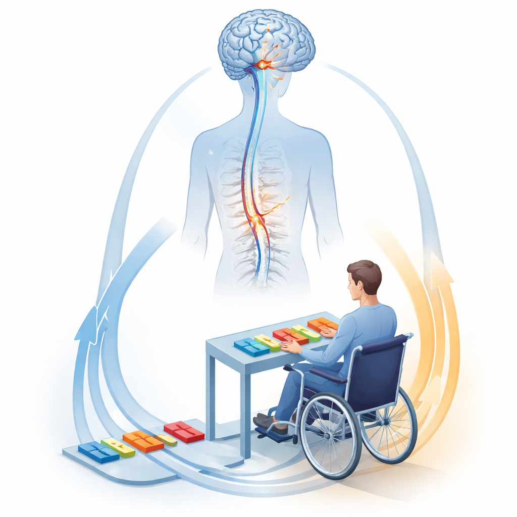
To probe how the nervous system adapts, the researchers enrolled 17 men with chronic spinal cord injury (more than six months, on average nearly eight years after injury) and 32 healthy men. Participants trained for four weeks on a computer-based rhythm game that required precise, timed movements of either the hands or the feet. In 60-minute supervised sessions, four times a week, they responded to arrows moving in time with beats, using either a tabletop device for the arms or a dance-mat-like platform for the legs. Performance was measured as how many cues they hit correctly and how closely they matched the ideal timing. At multiple points before, during, and after the training, everyone underwent detailed MRI scans designed to pick up tiny changes in brain structure.
Measuring hidden changes in brain tissue
The MRI methods went beyond traditional brain imaging. They included techniques that are sensitive to the amount and organization of brain tissue and to features related to myelin, the insulating sheath that helps nerve fibers send signals quickly. By following the same people over time, the team could track how gray matter (the brain’s processing centers) and white matter (the wiring that connects them) changed as training progressed. They focused on a network of regions known to be involved in learning new movements: the primary motor and sensory cortices, the cerebellum, the thalamus, the hippocampal formation, and the major pathways that carry signals from the brain to the spinal cord.
Performance gains and brain reshaping
Every participant with spinal cord injury improved over the month of training. They became both more accurate and faster, with gains leveling off after about one month and remaining stable when tested again nearly two months after training had stopped. At baseline, patients performed worse than healthy trainees, but during training they often showed larger overall improvements. MRI revealed that these behavioral gains were accompanied by widespread structural changes in both gray and white matter. The motor cortex, the long tracts that descend through the brainstem toward the spinal cord, and the cerebellum all showed time-dependent shifts in volume and in markers linked to myelin and fiber organization. Early in training, some areas briefly expanded and then partially shrank back, while measures tied to myelin and fiber alignment gradually strengthened over the full training period and stayed stable at follow-up.
Linking brain changes to better movement
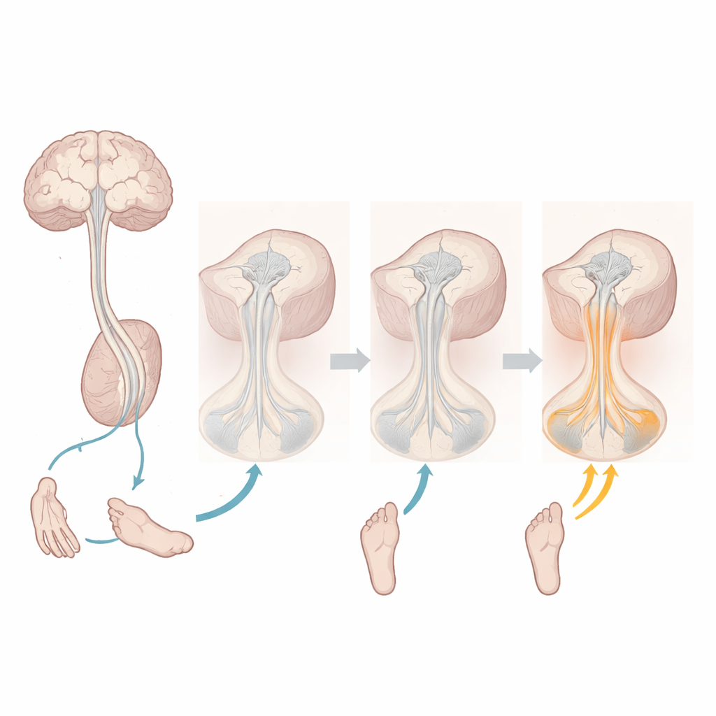
The patterns of remodeling were not random. Patients who showed bigger or faster improvements in the game tended to exhibit stronger structural changes in key movement pathways. For example, larger increases in tissue volume in the sensorimotor cortex were associated with quicker reaction-time gains, and specific changes along the corticospinal tracts—the main highways carrying movement commands—tracked with how quickly accuracy improved and how high it ultimately plateaued. The study also found body-specific effects: patients who trained with their legs showed more pronounced changes in leg-related regions of the motor system, whereas those who trained with their arms showed stronger shifts in arm-related areas of the brain, brainstem, and cerebellum. Strikingly, when spinal cord–injured trainees were compared directly with healthy trainees, the overall trajectories of brain plasticity were remarkably similar, with only minor differences in a few measures.
What this means for people living with spinal cord injury
To a non-specialist, the core message is hopeful: even years after a severe spinal cord injury, the brain still has a robust capacity to adapt to training. Intensive, engaging motor practice can reshape brain circuits important for movement, and those changes are closely tied to improvements in task performance. While this study does not yet prove that such training translates directly into everyday functional gains, it shows that the biological machinery for learning remains active long after injury. That insight supports the development of long-term, skill-based rehabilitation programs—possibly combined with other therapies—to harness this plasticity not just in spinal cord injury, but across many neurological conditions where recovery has traditionally been viewed as limited.
Citation: Emmenegger, T.M., David, G., Mohammadi, S. et al. Neuronal plasticity during motor rehabilitation training after spinal cord injury. Commun Biol 9, 561 (2026). https://doi.org/10.1038/s42003-026-09793-7
Keywords: spinal cord injury, brain plasticity, motor rehabilitation, neuroimaging, motor learning