Clear Sky Science · en
Molecular tension indicators reveal unexpectedly complex regulation of tension in live mouse organs
Seeing Invisible Forces Inside the Body
Every second, tiny mechanical forces tug on the molecules that hold our cells together and keep our organs in shape. These forces guide how an embryo forms, how the heart beats, and how tissues fail in disease—yet they are almost impossible to see. This study introduces a new way to watch those hidden pulls and stretches in living mouse organs, revealing that the internal “tension landscape” of our tissues is far more complex and finely tuned than previously thought.
A New Way to Watch Cellular Tug-of-War
For years, researchers have relied on specialized paired dyes to sense molecular tension, a method called FRET. While powerful, FRET-based sensors are hard to use deep inside real tissues because they are sensitive to optical noise and require careful calibration. The authors instead redesigned a single green fluorescent protein so that it subtly changes brightness when it is stretched. They inserted this flexible module into well-studied structural proteins, then attached a red fluorescent tag at the end. Because the green signal fades under load while the red signal stays constant, changes in tension show up simply as shifts in color from yellow-green toward orange-red under the microscope.
Measuring Force One Molecule at a Time
To be sure this new sensor really responds to force, the team pulled on individual molecules with optical tweezers—tiny laser-based “tractor beams” that can stretch single proteins. They attached the sensor to DNA handles, grabbed each end with microscopic beads, and increased the pulling force while monitoring green fluorescence. As the force rose from zero to a few trillionths of a newton, the sensor’s brightness changed in a predictable and reversible way. Within cells grown in dishes, drugs that relax the cell’s internal motors made the sensors glow greener, confirming that the tool faithfully reports changes in internal tension.
Different Structures, Different Force Patterns
The researchers next asked how tension varies within single cells. In one set of experiments, they followed forces on α-actinin, a protein that links actin filaments. They found that tension was higher near the bottom of the cell where it grips the surface and lower near the top, and that it changed over time in a restless, irregular fashion. Thin cell protrusions used for movement showed especially dynamic patterns: in broad, sheet-like edges, α-actinin tended to be more relaxed, while in finger-like projections, brief spikes of high tension appeared at both the tip and base, hinting at temporary anchor points that help cells explore their surroundings.
Hidden Force Maps in Heart and Liver
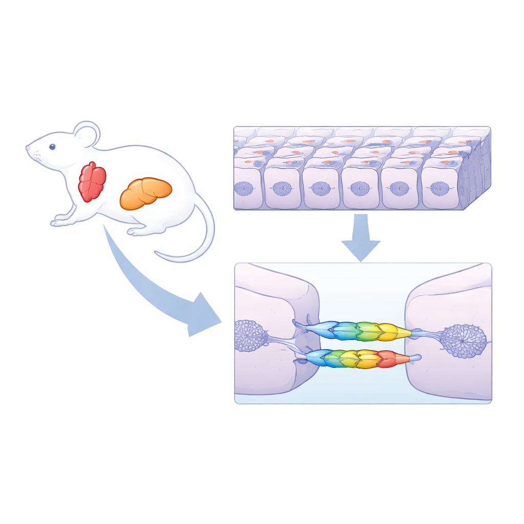
To see how these forces play out in real organs, the team created knock-in mice that produce the tension indicators in specific tissues. In heart muscle cells, α-actinin sits in the Z-discs, the regular stripes that organize contractile fibers. Superresolution imaging revealed a strikingly patchy pattern of tension along these stripes: even within a single band, some segments were under higher load than others. When the researchers relaxed the heart’s motor proteins with a drug, the Z-discs shifted uniformly toward the “relaxed” color, confirming that these patterns truly reflect mechanical strain. In the liver, they compared tension on α-actinin with that on α-catenin, a protein that links cell–cell junctions to the internal scaffold. Here, the two sensors painted very different maps: α-catenin was under high, nearly continuous tension along most cell borders, but was surprisingly relaxed around bile canaliculi and at special three-way junctions sealed by tight junctions. α-actinin, by contrast, showed a mosaic of high and low tension along the same borders.
Forces Are Shared in More Ways Than One
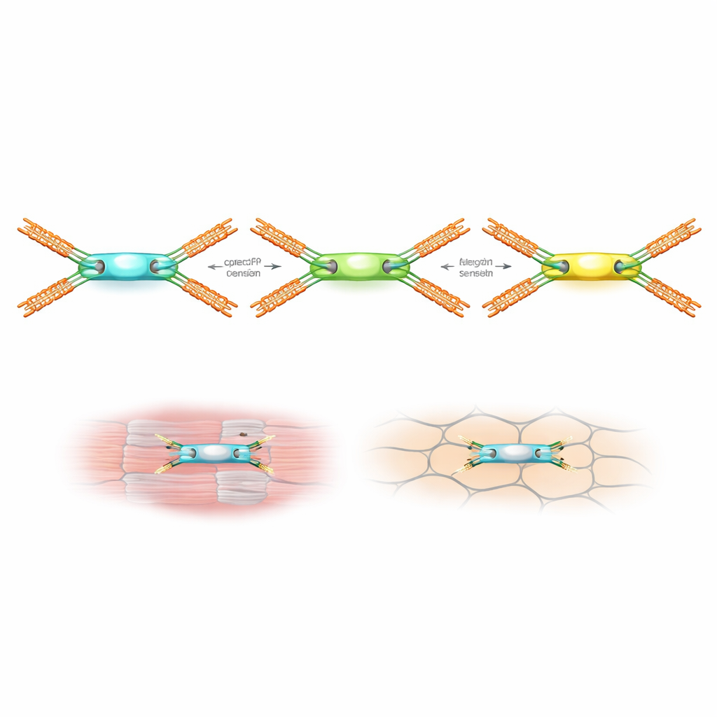
These findings suggest that tissues do not rely on a single “main” load-bearing molecule. Instead, different proteins can share, redistribute, or even avoid mechanical loads depending on local architecture and partners. Tight junctions in the liver, for example, appear to divert stress away from α-catenin at certain sites, while the same region still hosts patchy forces on α-actinin. In the heart, the fine-grained variations along Z-discs imply that even repetitive, rhythmic contraction is supported by a complex internal pattern of stress sharing. By offering a simple, high-resolution way to visualize such hidden force landscapes in living animals, these new molecular indicators open the door to studying how mechanical cues shape development, maintain organ function, and contribute to disease.
Citation: Fujiwara, K., Fujiki, K., Akama, T.O. et al. Molecular tension indicators reveal unexpectedly complex regulation of tension in live mouse organs. Commun Biol 9, 455 (2026). https://doi.org/10.1038/s42003-026-09746-0
Keywords: mechanobiology, molecular tension sensor, fluorescent protein, cell junctions, cardiac and liver tissue