Clear Sky Science · en
RoentMod: a synthetic chest X-ray modification model to identify and correct image interpretation model shortcuts
Why smarter X-ray AI matters
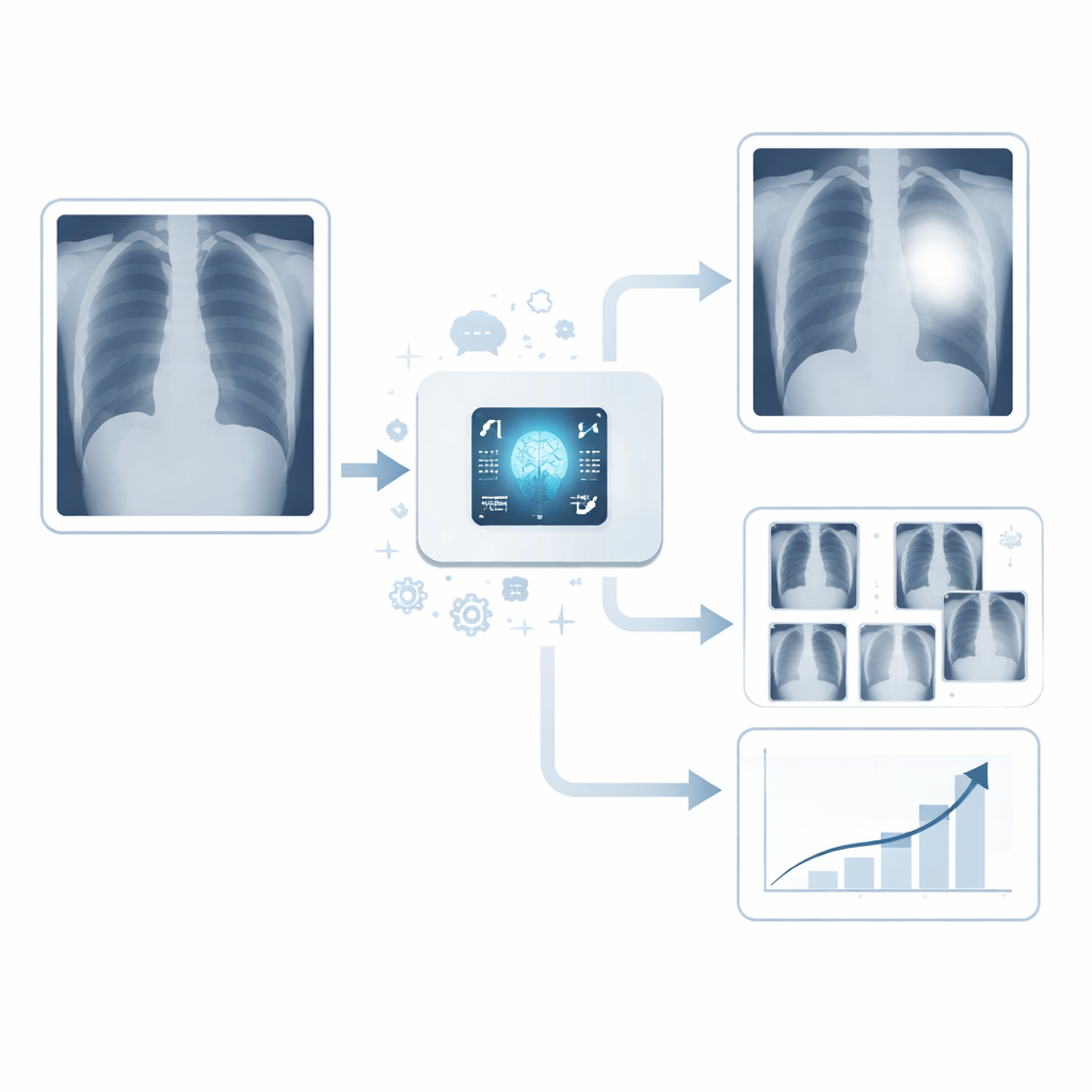
Chest X-rays are one of the most common medical tests in the world, used to spot problems with the heart, lungs, and chest. Computer programs powered by artificial intelligence (AI) can already read these images with impressive accuracy, promising faster diagnoses and less strain on radiologists. But these systems have a hidden weakness: they sometimes latch onto the wrong clues in an image—such as tubes, devices, or unrelated disease—as a shortcut instead of truly “looking” at the right finding. This paper introduces RoentMod, a new tool that creates realistic, modified chest X-ray images to uncover and fix these unreliable shortcuts in medical AI.
Making believable “what if” X-rays
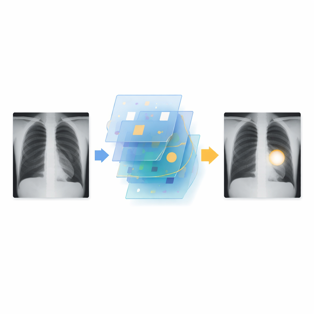
RoentMod is designed to answer a simple question: what would this same patient’s chest X-ray look like if they did—or did not—have a particular condition? The system starts from a real X-ray and a short text description, such as asking it to add fluid around the lungs or enlarge the heart. It then produces a new version of that same X-ray where only the requested change appears, while the rest of the anatomy stays the same. RoentMod builds on two existing image tools: one that knows how to generate realistic chest X-rays and one that can edit images based on text prompts. By reusing these components rather than training a new model from scratch, RoentMod can run quickly and on ordinary computer hardware.
Putting realism to the test
To see whether the edited images would fool experts, the researchers asked two radiologists to review 800 RoentMod-generated scans and additional mixed sets of real and synthetic images. In about 93% of cases, the modified images looked realistic, and unrequested extra problems appeared only rarely. For six common conditions—such as an enlarged heart, lung fluid, pneumonia, hernia, and lung masses—RoentMod successfully added the requested finding in nearly 9 out of 10 cases or better. The model was less reliable for subtler patterns like emphysema or tiny nodules, so those were excluded from later experiments. Image similarity tests and careful pixel-level checks showed that, aside from the edited region, the rest of the chest anatomy remained as consistent as in pairs of real X-rays taken from the same person at different times.
Revealing hidden shortcuts in existing AI
Armed with this controlled “what if” capability, the authors used RoentMod to stress-test several leading chest X-ray AI systems. They took scans that had no recorded disease, used RoentMod to add a single condition, and then observed how the models’ predictions changed for many different findings. Across all models, adding one disease often changed the predicted probabilities of other diseases that should have been unaffected—for example, adding fluid in the lungs could make the model more likely to predict a hernia. Saliency maps, which highlight the image areas the model relies on, showed that these shifts were not due to new signs of the other disease, but rather to the presence of any serious abnormality acting as a shortcut. Even powerful “foundation models” trained on huge datasets showed this behavior, although to a lesser degree.
Training AI to avoid easy but wrong answers
The team then flipped the script: instead of just testing models, they used RoentMod to help train a new one. They combined real chest X-rays from a large public collection with many RoentMod-edited versions in which exactly one chosen disease was added at a time. This exposed the model to carefully controlled examples where it could not safely assume that “sick” meant “everything is more likely.” When evaluated on several big chest X-ray datasets from different hospitals, the RoentMod-trained model showed better ability to distinguish specific diseases than a similar model trained only on real images. On internal tests, its performance improved by 3–19 percentage points, and it also outperformed the baseline on most diseases in outside datasets, though very large foundation models still led on some tasks.
What this means for future medical AI
For non-specialists, the takeaway is that RoentMod gives researchers a powerful, realistic way to ask targeted “what if” questions of medical AI systems. By editing real X-rays to add or remove specific findings while leaving everything else unchanged, RoentMod can reveal when models are taking misleading shortcuts and help retrain them to focus on the right signals. Although the current work centers on chest X-rays and a limited set of conditions, the same idea could extend to fairness checks across demographic groups, to other imaging types such as CT or MRI, and to AI systems that generate full radiology reports. In short, RoentMod shows that carefully crafted synthetic images can make medical AI both more accurate and more trustworthy.
Citation: Cooke, L.H., Jung, M., Brendel, J.M. et al. RoentMod: a synthetic chest X-ray modification model to identify and correct image interpretation model shortcuts. npj Digit. Med. 9, 324 (2026). https://doi.org/10.1038/s41746-026-02497-6
Keywords: chest X-ray AI, synthetic medical images, shortcut learning, counterfactual imaging, radiology deep learning