Clear Sky Science · en
Cellular landscape and diagnostic value of TROP2 in cerebrospinal fluid of lung adenocarcinoma leptomeningeal metastases
Why the fluid around the brain matters
Lung cancer can sometimes spread to the delicate layers that cover the brain and spinal cord, a condition called leptomeningeal metastasis. When this happens, cancer cells float in the clear fluid that bathes the brain, called cerebrospinal fluid. These hidden cells are hard to detect and treat, yet they can quickly threaten thinking, movement, and life itself. This study takes a close, cell-by-cell look at that fluid and discovers a new way to spot the disease using a protein signal, offering hope for earlier and more reliable diagnosis.
Mapping the hidden cells in brain fluid
Using single-cell RNA sequencing, the researchers analyzed more than 49,000 individual cells from the cerebrospinal fluid of six people with lung adenocarcinoma that had spread to the meninges. This technique reads which genes are switched on in each cell, allowing the team to build a detailed map of all the cell types present. They found immune cells such as different kinds of T cells, natural killer cells, monocytes, and macrophages, along with circulating tumor cells shed from the cancer. A small group of tumor cells showed clear signs of active division, hinting that only a fraction of cells may be driving growth in this harsh, nutrient-poor environment. 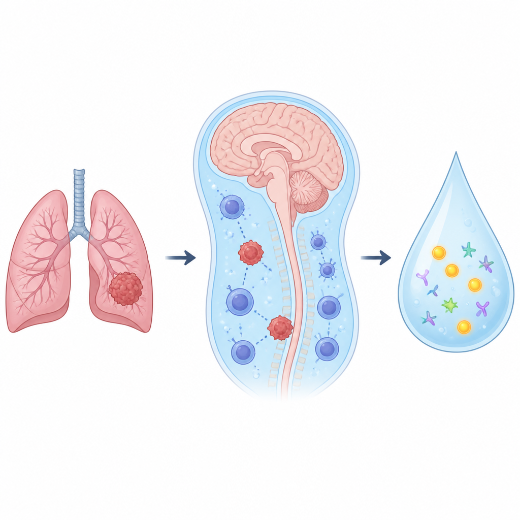
Immune defenses turned down
Although some T cells and natural killer cells in the fluid still carried molecules linked to killing cancer cells, the overall environment looked strongly immunosuppressive. In particular, the team identified specialized macrophages that resembled so-called wound healing or tumor-supporting cells. These macrophages expressed genes associated with dampening immune attacks and remodeling tissue, rather than fighting infection. Communication analyses showed that tumor cells sent inhibitory signals to T cells and natural killer cells through a checkpoint system centered on a receptor called TIGIT and its partner NECTIN2, and also used other pathways to tell immune cells not to eat or attack them. Together, these interactions painted a picture of cancer cells shaping the fluid into a safe harbor where they can survive and spread.
Cancer cells in fluid versus solid brain tumors
The researchers then compared tumor cells in the cerebrospinal fluid with tumor cells taken from solid brain metastases in the brain tissue itself. The two groups of cells shared some core gene regulators but also differed in important ways. Tumor cells in brain tissue had more active energy and growth pathways and stronger signatures of invasion and tissue remodeling, reflecting the richer, blood-fed brain environment. In contrast, fluid-based tumor cells appeared metabolically quieter, as if conserving resources. They also showed a distinct set of genes linked to stem-like behavior and poor outcomes in lung cancer, suggesting that a minority of highly adaptable cells may sustain disease in the fluid.
Finding a simple signal in the fluid
A key focus of the study was a cell-surface protein called TROP2, already known to be abundant on many solid tumors. The team found that circulating tumor cells in cerebrospinal fluid from lung cancer leptomeningeal metastasis patients strongly expressed TROP2. They then measured soluble TROP2 levels in the fluid of large patient groups. People with leptomeningeal metastasis from lung adenocarcinoma had TROP2 levels more than ten times higher than patients with lung cancer but no leptomeningeal spread or people without cancer. The test accurately separated those with and without leptomeningeal disease at a single cutoff level, and importantly, solid brain metastases alone did not raise TROP2 in the fluid. Similar patterns were observed in patients whose leptomeningeal metastases came from breast cancer. 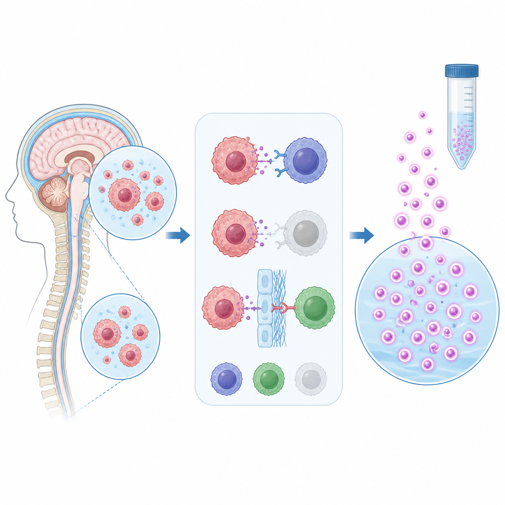
What this means for patients
For someone with lung cancer, diagnosing spread to the brain coverings currently relies on spotting cancer cells in a fluid sample or on subtle changes on brain scans, both of which can miss early or low-level disease. This work shows that the fluid environment becomes dominated by cancer-sparing immune signals and that a bloodless protein, TROP2, reliably flags when tumor cells have reached the cerebrospinal fluid. While more research is needed before routine use, measuring TROP2 in spinal fluid could become a practical laboratory test that helps doctors detect leptomeningeal metastasis sooner and distinguish it from other forms of brain involvement, guiding more timely and tailored care.
Citation: Wang, Z., Luo, J., Jin, Y. et al. Cellular landscape and diagnostic value of TROP2 in cerebrospinal fluid of lung adenocarcinoma leptomeningeal metastases. npj Precis. Onc. 10, 183 (2026). https://doi.org/10.1038/s41698-026-01379-0
Keywords: leptomeningeal metastasis, cerebrospinal fluid, lung adenocarcinoma, TROP2 biomarker, single-cell sequencing