Clear Sky Science · en
Maximizing pancreatic carcinoma classification performance using parrot optimized vision transformer
Why this matters for patients and doctors
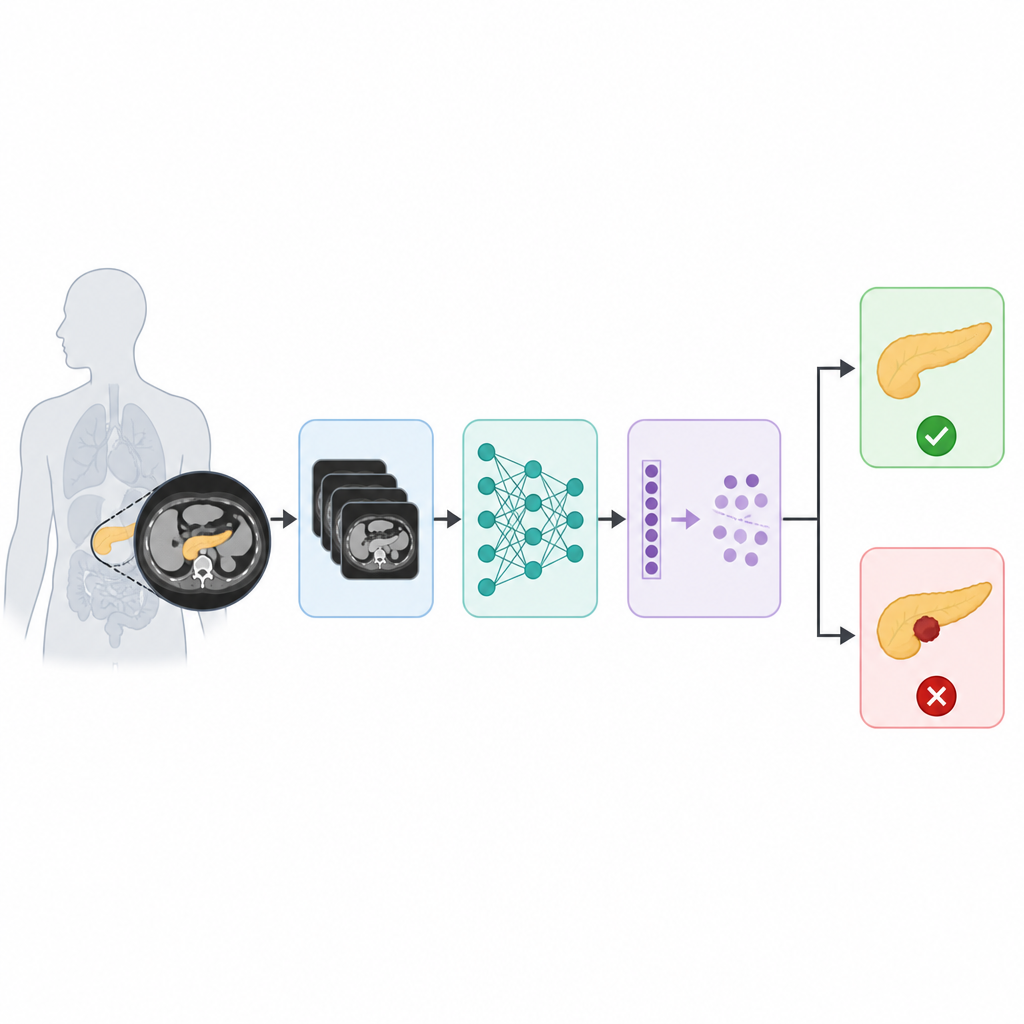
Pancreatic cancer is one of the deadliest cancers because it is usually found only after it has quietly grown for a long time. This study explores how a carefully designed artificial intelligence system can read CT scans of the abdomen and help doctors spot pancreatic tumors more reliably and consistently. By combining several modern image processing tools into one pipeline, the researchers show that computers can support earlier and more accurate diagnosis, which is crucial for improving the chances of successful treatment.
Turning raw scans into clearer pictures
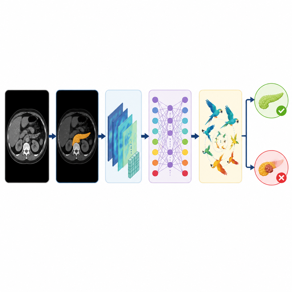
The journey begins with a collection of 1811 CT images of the pancreas, taken from an open online dataset and labeled as either normal or tumor. Because medical images can be noisy and slightly different from one scan to another, the team first increased and cleaned the data. They created extra training examples by rotating, flipping, and scaling the original images, which helps the system learn to cope with natural variation between patients and scanners. A special type of filter that is sensitive to edges and textures is then applied to sharpen important details in the pancreas while reducing distracting visual clutter. This careful preparation makes it easier for later steps to focus on subtle changes that may signal a tumor.
Finding the organ before judging its health
Rather than asking the computer to understand the whole CT slice at once, the researchers first teach it to find and outline the pancreas itself. They use a tried and tested segmentation network, shaped like a U, that learns to separate the organ from the surrounding organs and tissues in the abdomen. Once the pancreas region is isolated, this focused cutout is fed into a powerful detector that is skilled at spotting objects of different sizes. This detector translates the pancreas into rich numerical features that capture its shape, texture, and internal patterns at several scales, while ignoring most of the background. By narrowing attention in this way, the system reduces confusion from other structures that share the same image.
Letting an attention-based model decide
The distilled features then pass to a vision transformer, a newer kind of image model that originally came from language translation research. Instead of scanning the image piece by piece with sliding filters, this model looks at the pancreas in small patches and learns how each patch relates to every other patch. In simple terms, it pays attention to how patterns in one part of the organ connect to patterns elsewhere, which helps it capture both local details and overall context. The transformer produces an initial decision on whether the pancreas looks normal or cancerous. To push performance even further, the authors add a final refinement step inspired by the group behavior of parrots searching for food, where many candidate solutions are explored and gradually improved to reduce mistakes.
Putting the pipeline to the test
The complete system, which the authors call ViT-PO, is evaluated with standard measures that matter in medicine: how often it is right overall, how often it correctly flags tumors, and how often it avoids raising false alarms. On the test images, the model reaches about 99 percent overall accuracy and shows a strong balance between catching true tumor cases and not mislabeling healthy scans. It also performs better than several well-known alternatives, including classic machine learning methods, standard deep neural networks, and other transformer-based designs that do not use the same integrated pipeline. Cross-checks with different data splits and repeated runs suggest that the behavior of the system is stable and not just a lucky result on one particular subset.
What this means for future care
For a lay reader, the key takeaway is that combining several smart steps—cleaning the image, isolating the organ, describing it in detail, judging it with an attention-based model, and then fine-tuning the decision—can make computer support for pancreatic cancer diagnosis both more accurate and more reliable. While this work still uses a single dataset and focuses on a simple choice between normal and tumor, it points toward tools that could one day assist radiologists in catching pancreatic cancer earlier and with greater confidence, provided that they are carefully validated on larger and more diverse patient populations.
Citation: Mallika, C., Dinesh, E., Alsolai, H. et al. Maximizing pancreatic carcinoma classification performance using parrot optimized vision transformer. Sci Rep 16, 16277 (2026). https://doi.org/10.1038/s41598-026-53240-w
Keywords: pancreatic cancer, CT imaging, deep learning, vision transformer, medical diagnosis