Clear Sky Science · en
Microglia-specificity of different markers is overridden in glioblastoma specimens
Why this matters for brain cancer
Doctors and researchers are trying to harness the brain’s own immune cells to fight deadly tumors like glioblastoma. But inside a tumor, two closely related cell types mix together: resident brain defenders called microglia and wandering immune cells called macrophages. Many new therapies depend on telling these cells apart. This study shows that several of the most popular “ID tags” used to mark microglia stop being reliable inside glioblastoma, a warning that could reshape how scientists interpret brain tumor data and design treatments.
Guardians of the brain
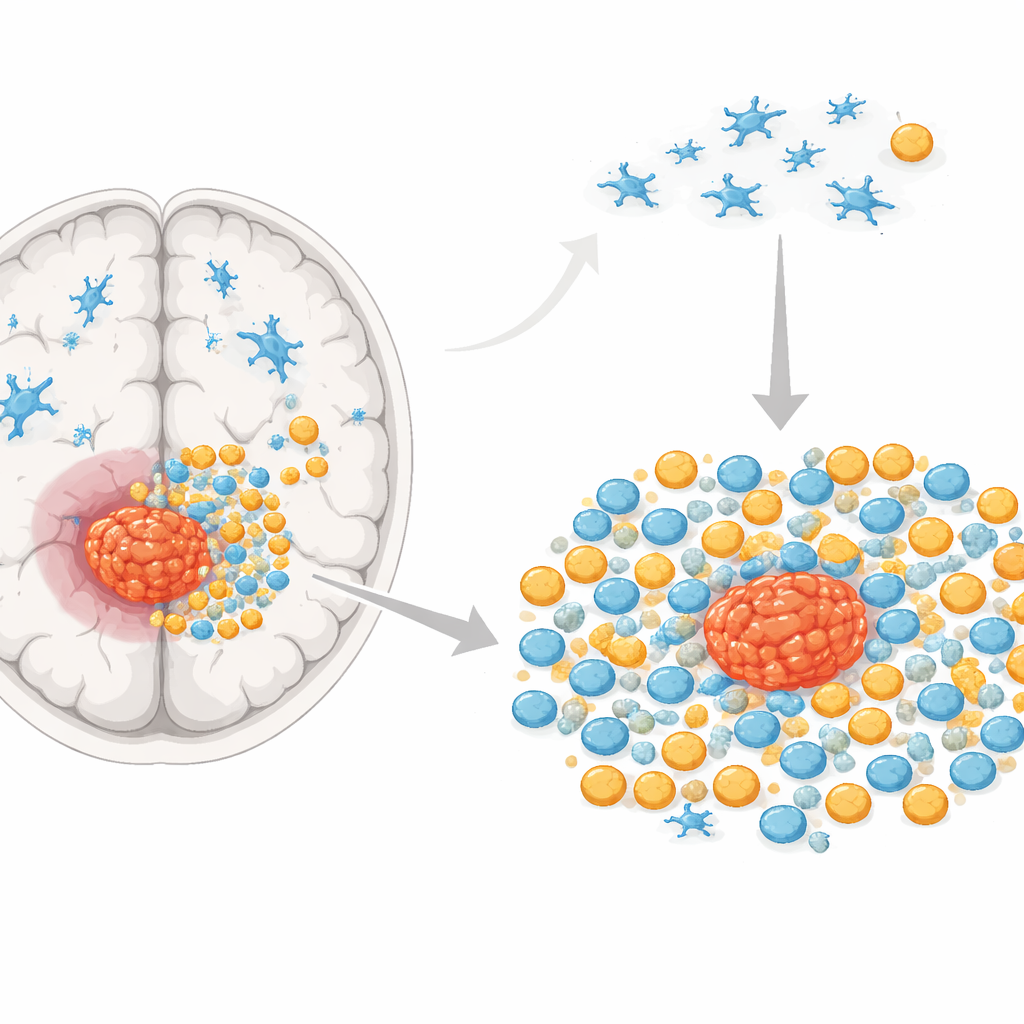
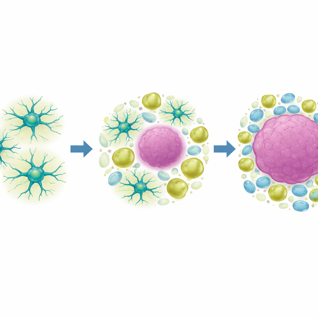
In a healthy brain, microglia quietly patrol the tissue, cleaning up debris and responding to injury. Macrophages, by contrast, come from the bone marrow and normally live elsewhere in the body, but can flood into the brain when there is damage or disease. In glioblastoma, these two cell types can make up as much as half of the tumor mass. Some may help the tumor grow and evade therapy, while others might restrain it. To understand their roles, researchers need clear, practical ways to distinguish microglia from macrophages in real tissue, not just in gene lists.
Markers that work in healthy brain
Over the past decade, powerful single-cell methods suggested that certain molecules are made almost exclusively by microglia. Four in particular—SALL1, TMEM119, P2RY12, and HEXB—have become widely used “microglia-only” markers in research. The authors tested whether these markers really behave that way at the protein level in human and mouse brain sections, using fluorescent staining. In tissue from people with epilepsy and from healthy mice, three of the four markers (SALL1, TMEM119, and P2RY12) were indeed found on the vast majority of microglia-like cells, while HEXB was less consistent. This supported the idea that, under almost normal conditions, these tags can identify microglia reasonably well.
How tumors blur cell identities
The situation changed dramatically in a mouse model of glioblastoma. When tumor cells were implanted into the brain, the team mapped where the markers were expressed: in tissue far from the tumor, at its edge, and deep within the tumor core. All four markers were present around and inside the tumor, but their pattern shifted. SALL1, TMEM119, and P2RY12 were strongly expressed near the tumor border and dropped sharply inside the tumor mass, leaving many immune cells without these supposedly defining microglial tags. HEXB was weak and patchy in all regions. The staining also appeared on cells that did not carry a general myeloid marker, hinting that some tumor cells themselves can express these molecules.
Macrophages pick up “microglia-only” tags
To prove which cells were which, the researchers created bone marrow chimeric mice, a technique that labels all incoming macrophages with a fluorescent signal while leaving resident microglia unlabeled. In these animals, they could directly see microglia and macrophages side by side in the same tumor. Contrary to expectations, both populations expressed SALL1, TMEM119, and P2RY12 in similar patterns, especially outside the tumor core. Inside the tumor, only a minority of either cell type carried these markers. In dishes, primary microglia and bone‑marrow–derived macrophages both produced all four markers, and a microglial cell line exposed to tumor-conditioned fluid reduced its levels of SALL1, TMEM119, and P2RY12—mirroring the loss of marker signal seen in tumors.
Rethinking microglia ID in cancer
Taken together, the findings show that these four molecules cannot be treated as exclusive microglia identifiers in the context of glioblastoma. Tumor conditions cause microglia to lose the markers, and macrophages and even tumor cells can gain them, effectively erasing the clean boundaries suggested by earlier gene-expression studies. For lay readers, the message is that in brain cancer, immune cells are highly adaptable and change their “surface badges” in response to the tumor environment. Researchers will need to use more complex, context-aware strategies—such as fate-mapping models or combinations of markers—rather than relying on any single label to distinguish microglia from macrophages when developing and testing new glioblastoma therapies.
Citation: Bungert, A.D., Sanchin, A., Blank, A. et al. Microglia-specificity of different markers is overridden in glioblastoma specimens. Sci Rep 16, 14687 (2026). https://doi.org/10.1038/s41598-026-52315-y
Keywords: glioblastoma, microglia, macrophages, brain tumor immunity, cell markers