Clear Sky Science · en
RDE-DR: robust deep ensemble CNNs for automated diabetic retinopathy detection from fundus images
Why eye scans matter for people with diabetes
Diabetic retinopathy is a complication of diabetes that slowly damages the light‑sensing tissue at the back of the eye. Caught early, it can often be treated before vision is lost. But checking thousands of retinal photographs by hand is time‑consuming for specialists. This study explores how a carefully designed mix of artificial intelligence models can help screen these images more reliably, so that people at risk are flagged sooner and healthy patients avoid unnecessary follow‑ups.
Looking for warning signs in eye photographs
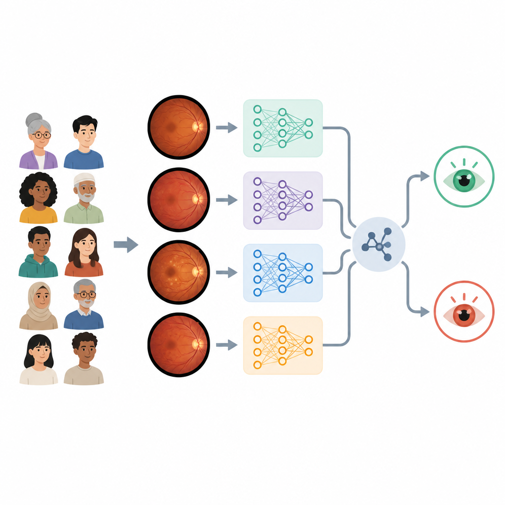
The researchers focus on color photographs of the retina, known as fundus images, from a public collection called the APTOS 2019 dataset. These pictures show tiny blood vessels and spots of bleeding or leakage that signal diabetic damage. The team converts the original five medical grades into a simpler question that matters for large‑scale screening: does this eye show any diabetic retinopathy or not. This turns the task into a yes or no decision that an automated system could make rapidly for thousands of patients.
Making hidden details easier for computers to see
Real‑world eye photographs vary a lot in sharpness, brightness, and color, which can confuse computer models. To reduce this problem, the authors use a contrast‑boosting technique called CLAHE that brightens local details without exaggerating noise. They resize all images to a standard small square, normalize the colors, and use random rotations and flips during training so the system does not overfit to a single camera angle or lighting condition. The dataset is split so that four fifths of the images teach the models and one fifth is held back to test how well the system generalizes to new cases.
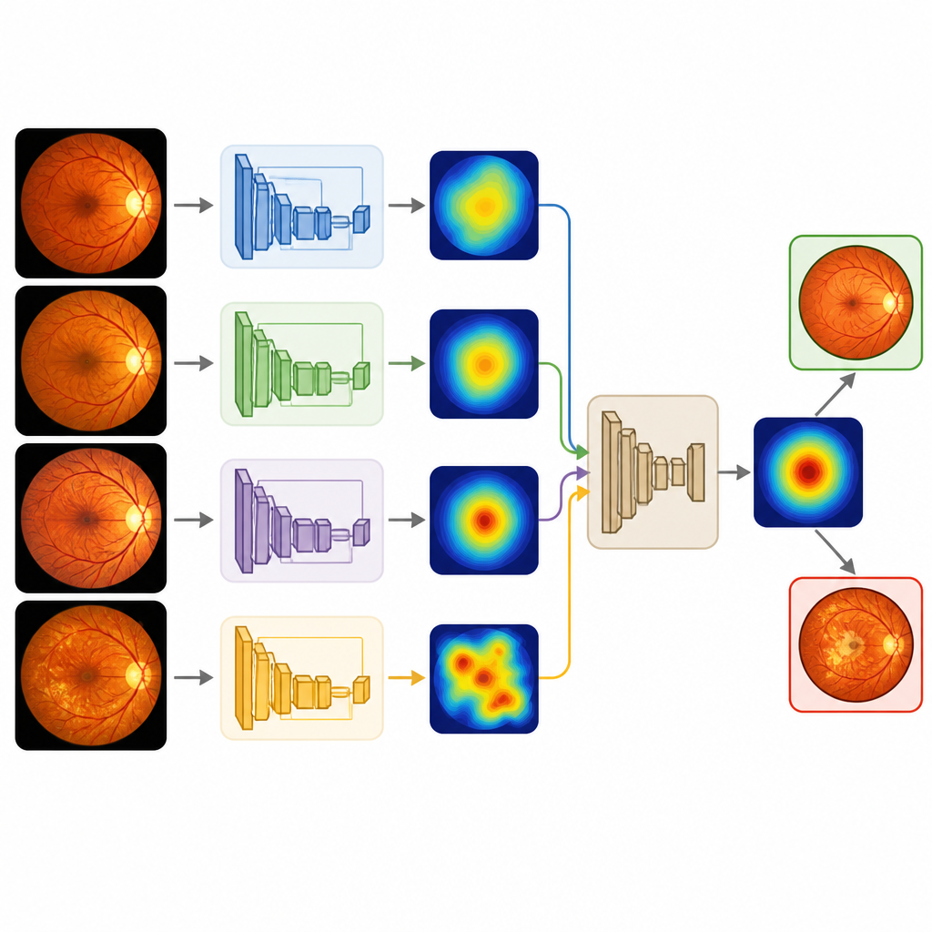
Many eyes instead of one
Rather than depending on a single deep learning model, the study trains four popular convolutional neural networks that were first developed on a large everyday photo collection. These models, known in the field as VGG16, VGG19, ResNet50, and DenseNet121, are retrained to recognize healthy and diseased retinas. Each one already performs very well on its own, correctly classifying about 98 percent of test images and missing only a handful of diseased eyes. The key idea of the work is to combine their strengths through several decision‑fusion schemes, including simple voting, average confidence, and more advanced fuzzy logic‑inspired rules.
Combining model opinions with care
The team systematically studies seven ways of blending the four model outputs, always under the same training recipe. They also fine‑tune the cut‑off point where a probability is turned into a yes or no prediction, rather than assuming a fixed halfway mark. By scanning through many thresholds, they measure how accuracy, sensitivity, and precision trade off, which is crucial for screening tools that must avoid both missed disease and excessive false alarms. They then look beyond simple accuracy, examining how confidently the models separate healthy from diseased images using receiver‑operating curves and probability density plots.
What the results mean for future eye screening
Across the board, the ensemble methods maintain or slightly improve on the already strong single‑model results, reaching about 98.6 percent accuracy and a very high score for separating classes. Simple schemes like majority voting or averaging confidence values are remarkably stable, while one of the fuzzy methods proves more sensitive to design choices and yields less balanced performance. For a layperson, the main message is that combining several well‑trained image readers, after carefully cleaning the pictures and tuning how their votes are merged, leads to a more trustworthy automated assistant. Such a tool will not replace eye doctors but could help them quickly sort large numbers of retinal photographs, focusing precious clinic time on the patients whose eyes show early signs of trouble.
Citation: Aiche, I., Brik, Y., Attallah, B. et al. RDE-DR: robust deep ensemble CNNs for automated diabetic retinopathy detection from fundus images. Sci Rep 16, 15226 (2026). https://doi.org/10.1038/s41598-026-48669-y
Keywords: diabetic retinopathy, fundus imaging, deep learning, ensemble models, medical screening