Clear Sky Science · en
Feasibility of automated AI-based contouring and stable radiomic feature assessment by HyperSight-CBCT Imaging for adaptive high-precision radiotherapy of prostate cancer
Faster, Smarter Cancer Treatment Planning
For men with prostate cancer, modern radiation therapy can be remarkably precise, but that precision comes at a cost: doctors must painstakingly trace the prostate and nearby organs on many imaging slices before each treatment plan is ready. This study asks a timely question: can artificial intelligence safely take over most of that contouring work on a new, high-quality cone‑beam CT scanner, while still providing reliable image-based measurements that might one day guide fully personalized treatment?
Seeing Inside the Pelvis in Real Time
Today’s high-precision radiotherapy often uses a planning CT scan taken before treatment and additional scans taken right on the treatment machine, just before each dose. The newer HyperSight cone‑beam CT system can quickly capture detailed images of the pelvis with lower radiation dose, making it attractive for adaptive radiotherapy—where the plan is adjusted to the anatomy of the day. In this study, 50 men with prostate cancer underwent both standard planning CT and HyperSight scans. The researchers focused on several key pelvic structures: the prostate, bladder, rectum, seminal vesicles, penile bulb, and both hip joints.
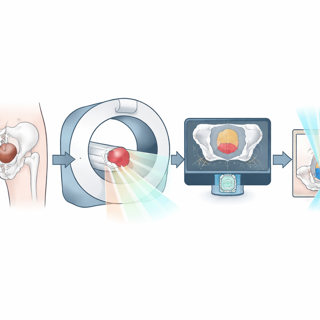
Letting the Computer Draw the Lines
The team compared three ways of outlining organs on the images. In the fully manual approach, physicians drew all boundaries themselves. In the AI-only mode, a trained algorithm automatically created the contours. A hybrid mode started with AI contours that physicians then checked and refined. For each method, the researchers measured how closely different contours matched, how far their edges differed, and how much the organ centers shifted. They also timed how long each approach took. For large, clearly visible structures such as the bladder and hip bones, agreement between all methods was very high. For the prostate and rectum it was slightly lower, but still strong. Small or fuzzy structures like the seminal vesicles and penile bulb proved more challenging for every method, including AI.
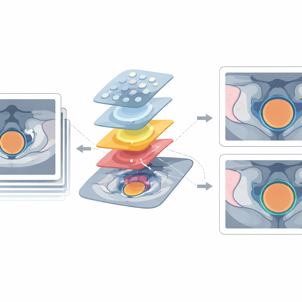
From Pictures to Quantitative Fingerprints
Beyond simple shape comparisons, the study examined “radiomic” features—hundreds of numbers extracted from the images that describe tissue brightness, texture, and shape. These features are increasingly explored as potential imaging biomarkers that might predict how tumors respond to treatment or which patients are at higher risk of side effects. The researchers tested how stable these numerical fingerprints remained when contours came from different methods and from different scan types. Overall, the radiomic features were highly consistent, especially for larger, high-contrast organs such as the bladder, prostate, rectum, and hip bones. Texture-based features, which describe patterns of pixel intensities, were particularly robust. Shape-based features and those from very small organs were more sensitive to small changes in the contours.
Saving Time Without Sacrificing Quality
Time is critical in adaptive radiotherapy: the longer it takes to contour and recalculate the plan, the greater the chance that the patient’s anatomy shifts between imaging and treatment. In this study, AI-only contouring reduced segmentation time by more than 90% compared with fully manual work, while the hybrid AI-plus-physician approach still cut time by about 60%. Importantly, these gains were similar for both the planning CT and the HyperSight scans, and interobserver tests showed that using AI did not introduce extra disagreement between different physicians. This suggests that AI can make the workflow much more efficient while maintaining, and sometimes even improving, consistency.
What This Means for Patients
Put simply, the study shows that smart software can reliably outline the prostate and most nearby organs on modern cone‑beam CT images, and that the resulting image-based “fingerprints” of the tissue remain stable enough to be trusted in research and, potentially, in future clinical decision-making. While small, hard‑to‑see structures still benefit from human fine‑tuning, AI-driven and hybrid contouring already offer high accuracy and substantial time savings. This opens the door to truly adaptive radiotherapy sessions where treatment can be quickly tailored to the anatomy and, one day, perhaps to radiomic markers that signal how each patient’s cancer is responding in real time.
Citation: Schmidt, R., Bajerski, D., Bicu, A.S. et al. Feasibility of automated AI-based contouring and stable radiomic feature assessment by HyperSight-CBCT Imaging for adaptive high-precision radiotherapy of prostate cancer. Sci Rep 16, 12191 (2026). https://doi.org/10.1038/s41598-026-46359-3
Keywords: prostate cancer radiotherapy, AI autocontouring, cone-beam CT imaging, radiomics, adaptive radiation therapy