Clear Sky Science · en
Glial cell mapping in the camel cerebellar cortex: a histochemical and immunohistochemical study
Why Camel Brains Matter
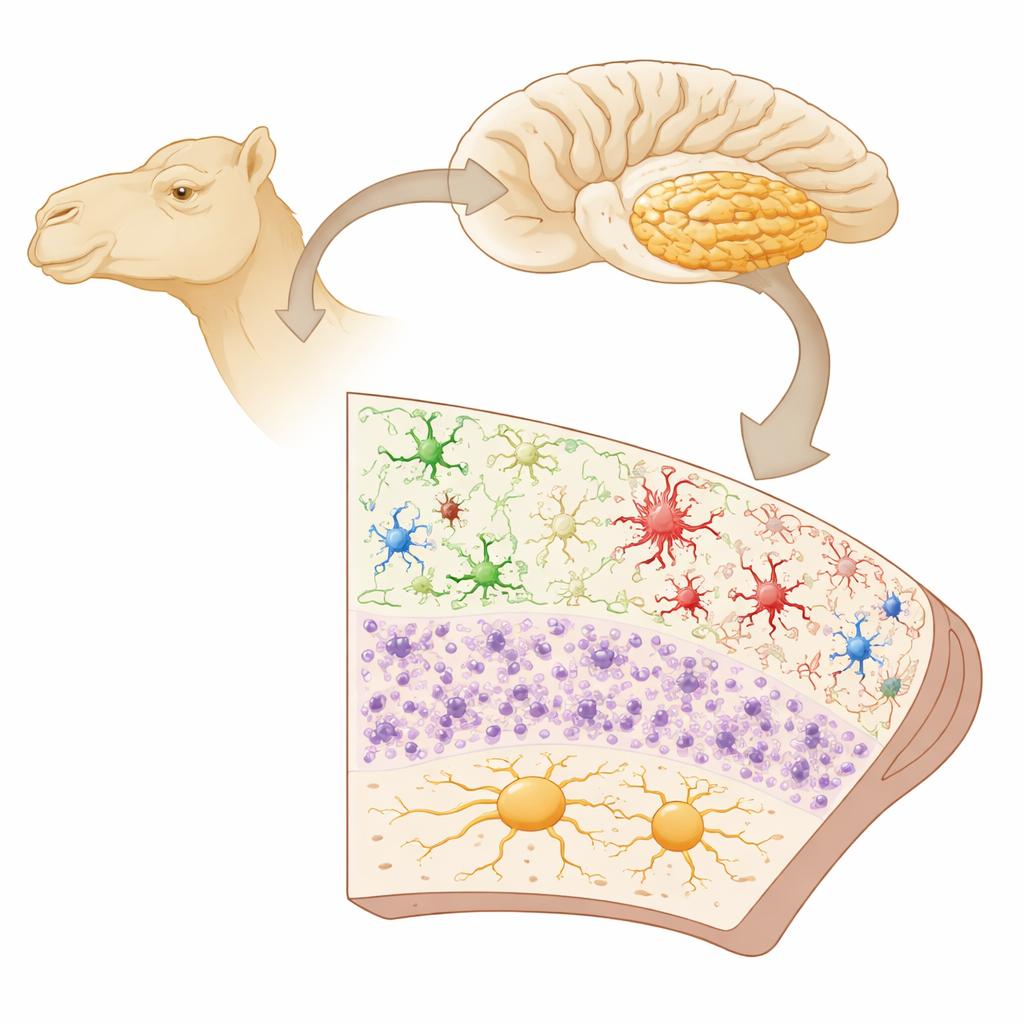
The brains of camels may seem like an unlikely frontier for neuroscience, but they offer a powerful window into how different mammals fine-tune balance, movement, and perhaps even resilience to harsh environments. This study takes a close look at the “little brain” at the back of the camel’s head—the cerebellum—and maps the support cells, called glia, that keep its nerve cells working smoothly. By comparing camel cerebellar glia to those of other species, the work helps scientists understand which features of brain wiring are shared across mammals and which are uniquely adapted.
The Little Brain Behind Precise Movement
The cerebellum coordinates smooth walking, precise limb control, eye movements, and even aspects of thinking and emotion. In all mammals, its outer sheet, the cerebellar cortex, has a layered architecture: an outer molecular layer, a middle sheet of large Purkinje cells, and an inner granular layer sitting on a core of white matter. While neurons transmit electrical signals, they are only one part of the story. Glial cells—astrocytes, oligodendrocytes, microglia, and a cerebellum-specific type called Bergmann glia—nourish neurons, insulate their fibers, maintain chemical balance, and patrol for damage or infection. Yet for camels, which are vital domestic animals in large parts of the world, these glial populations had barely been described.
How the Study Was Done
Researchers collected cerebella from ten healthy adult one-humped camels slaughtered for meat in Egypt. After carefully fixing and slicing the tissue, they used a combination of classic stains and antibody-based labeling to highlight different glial types. One marker (GFAP) revealed most astrocytes; S-100 identified Bergmann glia and fibrous astrocytes; Olig2 labeled oligodendrocytes, the cells that make insulating myelin; and Iba1 highlighted microglia, the brain’s resident immune cells. Light and electron microscopy allowed the team to examine cell shapes and their relationships to blood vessels and nerve fibers, while image-analysis software quantified how densely each cell type populated distinct cerebellar layers.
The Camel’s Glial Landscape
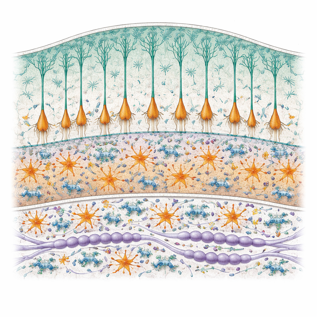
The team found that camel astrocytes have the familiar star-like appearance seen in other mammals, sending out fine processes that wrap around blood vessels and help form the blood–brain barrier—a cellular shield that tightly controls what enters brain tissue from the bloodstream. However, their distribution was strikingly uneven. Astrocytes were common in the granular layer and especially in the white matter, but a standard astrocyte marker (GFAP) revealed virtually none in the molecular layer, unlike in humans, monkeys, and rodents. This suggests that the astrocytes in that outer layer either use very low levels of this protein or rely on different molecular tools, hinting at species-specific specializations.
Specialized Support Cells in Layers
Bergmann glia, a specialized type of astrocyte unique to the cerebellum, formed 4–6 dense rows alongside Purkinje cells. Their long, straight processes ran like scaffolding from the Purkinje layer all the way through the molecular layer to the brain surface, creating vertical cables that likely guide connections and stabilize synapses. These cells were extremely numerous—over 5,000 per square millimeter—outnumbering Purkinje neurons. Oligodendrocytes were abundant in the white matter and also present in the granular layer, often lined up like beads along myelinated fibers, where they help maintain rapid signal conduction. Microglia showed remarkable variety: their shapes and orientations changed from layer to layer, and they were most densely packed in the white matter and granular layer, where they frequently contacted neurons, oligodendrocytes, and blood vessels or engulfed small, dying cell fragments.
What These Findings Tell Us
Taken together, the results show that camel cerebellar glia follow the same broad plan seen in other mammals—a three-layered cortex supported by astrocytes, Bergmann glia, oligodendrocytes, and microglia—yet display distinct patterns of density, shape, and molecular labeling. These differences may reflect evolutionary tuning of the camel’s motor system or unique responses to environmental stress, though functional tests are still needed. By providing a detailed cellular map, this work lays the groundwork for future studies on how camel brains cope with disease or injury and enriches the broader effort to understand how diverse mammalian species build and maintain a highly reliable “little brain.”
Citation: Attaai, A.H., Noreldin, A.E., Nomir, A.G. et al. Glial cell mapping in the camel cerebellar cortex: a histochemical and immunohistochemical study. Sci Rep 16, 13404 (2026). https://doi.org/10.1038/s41598-026-46231-4
Keywords: camel cerebellum, glial cells, astrocytes, microglia, oligodendrocytes