Clear Sky Science · en
Assessing bone radiodensity and thickness in cochlear implant patients through manual photon-counting CT image segmentation using ITK-SNAP
Why this matters for people with hearing loss
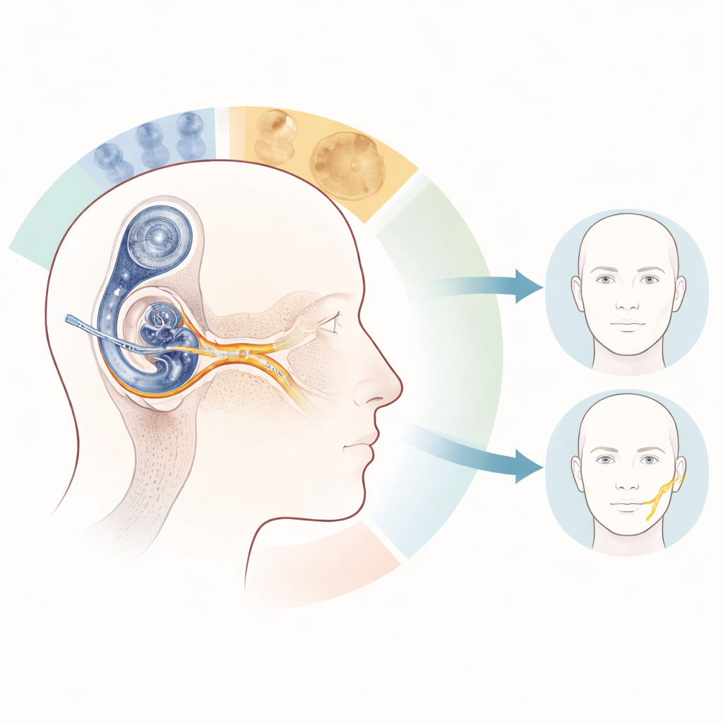
Cochlear implants have transformed life for many people with severe hearing loss, but in a small number of patients they can accidentally trigger the facial nerve, causing unwanted twitching or spasms on one side of the face. This study explores whether ultra-detailed CT scans can help doctors see the tiny strip of bone that separates the implant’s electrodes from the facial nerve, in hopes of better understanding and ultimately reducing this troubling side effect.
A closer look at the ear’s wiring
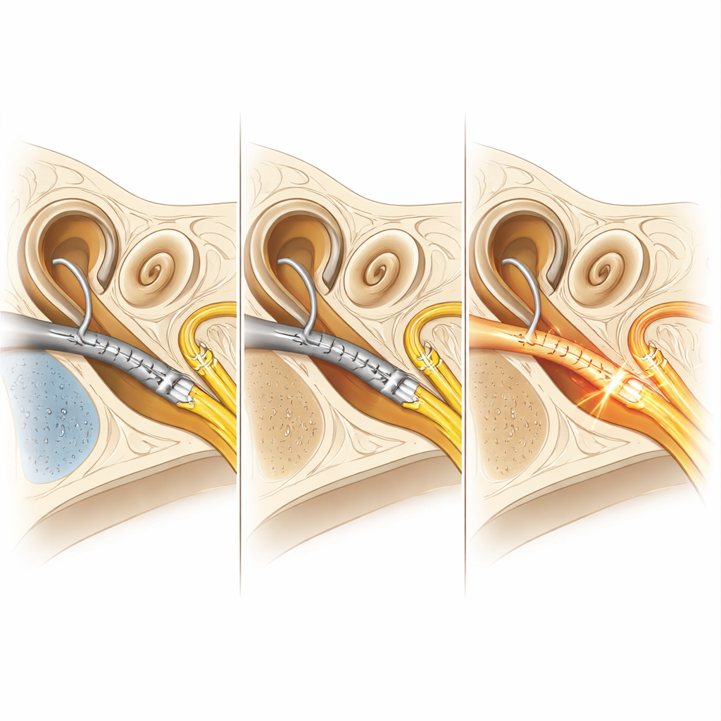
Inside the skull, the hearing nerve and facial nerve run very close together. A cochlear implant works by sending electrical signals along a flexible wire of electrodes inserted into the inner ear’s spiral-shaped cochlea. In rare cases, some of that electrical current can spill over and activate the nearby facial nerve instead of just the hearing nerve. This problem, called facial nerve stimulation, appears to be more common in people with a kind of abnormal bone growth in the ear called otosclerosis, which can change the structure and composition of the surrounding bone.
Using sharper scans to see tiny details
The researchers used a new type of CT scanner, called photon-counting CT, which produces very sharp images while using less radiation than conventional scanners. They combined these scans with ITK-SNAP, an open-source computer program that allows experts to manually draw and measure three-dimensional regions inside medical images. In this case, an ear surgeon carefully outlined the sliver of bone between the middle section of the cochlear implant’s electrode array and the nearby facial nerve, slice by slice, to calculate both how thick that bone was and how dense it appeared on the scan.
Comparing patients with and without facial twitching
The team studied nine adults with cochlear implants, dividing them into four groups: patients with facial nerve stimulation and severe otosclerosis, patients with facial nerve stimulation but no otosclerosis, and two small control groups without facial nerve problems, with and without otosclerosis. They compared hearing test results, surgical details, implant programming, bone thickness, and bone radiodensity between these groups. Overall hearing performance two years after implantation was similar across all groups, and standard implant settings such as electrode impedances and stimulation levels did not differ in a meaningful way.
What the bone measurements revealed
The ultra-detailed scans showed a clear difference in bone properties linked to otosclerosis itself. Patients with far advanced otosclerosis had substantially lower bone radiodensity around the region between the cochlea and facial nerve than patients without otosclerosis, confirming that their bone is genuinely different in quality. However, when the researchers compared patients who did and did not experience facial nerve stimulation, they did not find a consistent difference in either bone density or the average thickness of the bony bridge between the implant and the nerve. One striking exception was a patient without otosclerosis whose bone layer was extremely thin—only about a tenth of a millimeter—suggesting that in some individuals, simple physical closeness may be enough to let current “leak” to the facial nerve.
What this means for future cochlear implant care
For a layperson, the key message is that this new scanning and measurement approach can reliably show just how solid and how thick the bone is between a cochlear implant and the facial nerve, and it confirms that otosclerosis really does soften that bone. But lower bone density alone does not fully explain why some people develop facial twitching and others do not. Instead, an extra-thin strip of bone in a few patients, along with individual differences in implant programming and anatomy, may tip the balance. This small, early study shows that photon-counting CT combined with precise manual image analysis is a promising way to study these questions and could eventually help surgeons and audiologists better predict and prevent facial nerve side effects in cochlear implant users.
Citation: Quatre, R., Bonnard, Å., Eklöf, M. et al. Assessing bone radiodensity and thickness in cochlear implant patients through manual photon-counting CT image segmentation using ITK-SNAP. Sci Rep 16, 13403 (2026). https://doi.org/10.1038/s41598-026-45916-0
Keywords: cochlear implant, facial nerve stimulation, otosclerosis, photon-counting CT, bone density