Clear Sky Science · en
Ultra-widefield OCTA assessment of retinal and choroidal microcirculation in cerebral small vessel disease
A New Way to Look at Hidden Brain Vessel Damage
Cerebral small vessel disease quietly harms tiny blood vessels deep inside the brain, raising the risk of stroke and memory problems. Because these vessels are too small to see directly, doctors have struggled to spot early changes before lasting damage occurs. This study explores whether detailed scans of the back of the eye can act as a simple window into brain vessel health, offering a noninvasive way to track this common but often overlooked condition.
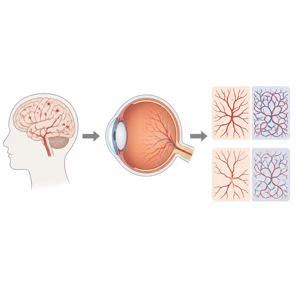
The Eye as a Window into the Brain
The blood vessels of the eye and brain develop from the same source and share similar structure and function. That connection has long tempted scientists to use the eye as a stand in for the brain. Modern imaging now allows doctors to map tiny vessels in the retina and a deeper layer called the choroid, which feeds the light sensitive tissue. In this work, researchers used ultra widefield optical scans that capture a very large area at the back of the eye, far beyond the central region usually examined. Their goal was to see whether subtle differences in these vessels could reveal the presence and severity of small vessel disease in the brain.
How the Study Was Carried Out
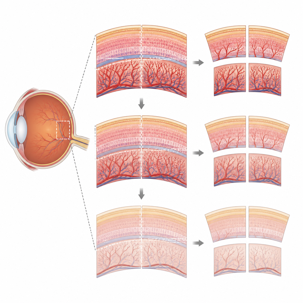
The team examined 40 people with cerebral small vessel disease and 30 similar but healthy volunteers. All participants underwent brain MRI scans, memory testing, and detailed eye exams. The MRI images were used to assign each patient a burden score based on common signs of small vessel damage, such as tiny strokes, white matter changes, and small bleeds. For the eye, the researchers used swept source optical coherence tomography angiography, a fast, noninvasive scan that builds three dimensional maps of the retina and choroid. They divided the back of the eye into nine regions and measured overall vessel density, the thickness of the choroid, and how much of its volume was made up of blood vessels versus supporting tissue.
Where the Eye Shows the Strongest Signals
The most striking changes appeared in the nasal and inferior regions of the eye, the areas closer to the nose and lower part of the visual field. In people with small vessel disease, these regions showed a thinner choroid and a lower share of space occupied by blood vessels, meaning both the outer coat and its blood supply were reduced. The fine vessel networks in the retina above these regions were also less dense. When the researchers adjusted for age, blood pressure, and sex, these links remained strong. As the MRI burden score increased, the loss of choroidal tissue and vessels spread from a few regions to almost the entire area imaged, suggesting a stepwise worsening that mirrored disease severity in the brain.
Differences Across Age Groups
Because small vessel disease is more common in older adults, the team checked whether age changed the pattern of eye findings. They split patients into younger and older groups and repeated their analysis. In younger patients, the strongest links between eye measures and disease were confined mainly to a few nasal regions. In older patients, both choroidal thinning and reduced vessel share were tied to disease across many more regions of the eye. This widening pattern with age supports the idea that ongoing small vessel damage gradually reshapes the choroidal circulation over time.
What This Means for Patients and Doctors
For a layperson, the key message is that very detailed pictures of the blood supply behind the retina can reflect what is happening in the tiny vessels of the brain. In this study, especially in the nasal and lower parts of the eye, a thinner and less vessel rich choroid was linked to higher small vessel disease burden on MRI. While the work does not yet prove cause and effect, it suggests that a quick, noninvasive eye scan could one day help doctors screen for early brain vessel damage, follow its progression, and perhaps guide efforts to protect brain health before strokes or memory decline appear.
Citation: Zhou, Y., Gao, C., Zhang, X. et al. Ultra-widefield OCTA assessment of retinal and choroidal microcirculation in cerebral small vessel disease. Sci Rep 16, 14964 (2026). https://doi.org/10.1038/s41598-026-45896-1
Keywords: cerebral small vessel disease, retinal imaging, choroid, OCTA, brain microcirculation