Clear Sky Science · en
A hybrid dual-stream CNN framework with dynamic data augmentation and improved Manta Ray Foraging Optimization for robust glaucoma detection
Why this research matters for your vision
Glaucoma is a silent thief of sight: it slowly damages the optic nerve without obvious symptoms until vision loss is often permanent. Regular eye exams can catch it early, but reading retinal images accurately takes time, expensive equipment, and expert clinicians. This paper presents an artificial intelligence (AI) system designed to act as a fast, reliable assistant for doctors, spotting glaucoma in common eye photographs with accuracy that rivals, and in some tests even surpasses, current methods.
The problem of a quiet eye disease
Glaucoma affects tens of millions of people worldwide and is a leading cause of irreversible blindness. The disease gradually alters the appearance of the optic nerve head—the point where nerve fibers exit the eye—changing the relative size and shape of the central “cup” inside the optic disc and the surrounding nerve tissue. Today, clinicians rely on a mix of tests, including retinal photographs, visual field exams, and pressure measurements inside the eye. These methods work, but they are labor‑intensive, depend heavily on specialist interpretation, and can be inconsistent from one clinic or observer to another. Meanwhile, large numbers of eye images are now captured in routine care, creating a natural opportunity for automated tools that could flag risky cases early.
Teaching a computer to read eye photographs
To build such a tool, the authors start with color fundus photographs—standard images of the back of the eye—from four public glaucoma datasets. They first run each image through a careful cleaning pipeline: noise is smoothed, brightness and contrast are standardized, and distracting black borders are removed. A clustering method then singles out the region around the optic disc and cup, which is cropped and resized so that every image presents the most informative area in a consistent way. This step ensures that the AI focuses on the structures eye doctors actually use to judge glaucoma, rather than irrelevant background details.
Making the most of limited and messy data
A major challenge in medical imaging is that truly diseased cases are scarce compared with normal ones, and real‑world images vary in sharpness, lighting, and even camera type. To address this, the authors design a “hybrid data augmentation” strategy. They create realistic variations of each image by rotating, shifting, zooming, and flipping it, much like taking the same eye photo from slightly different positions. On top of that, they inject a tailored amount of gentle visual noise, calculated from each image’s brightness and contrast, to mimic the imperfections of everyday clinical imaging. This controlled variety helps the AI learn to recognize glaucoma across a broad range of conditions instead of overfitting to a narrow, idealized dataset. 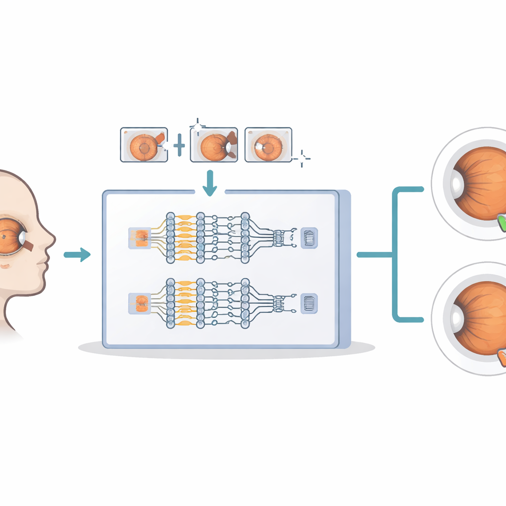
Two expert viewpoints and a focused gaze
At the heart of the system is a dual‑stream deep learning design that behaves like two complementary specialists examining the same eye. One stream, based on a network called DenseNet121, concentrates on large‑scale structure: the outline of the optic disc, the shape and size of the cup, and overall deformation of the tissue. The other stream, based on ResNet50, dwells on fine texture: subtle patterns in the nerve fiber layer and small irregularities that can signal early damage. A lightweight “attention” mechanism then acts like a spotlight, automatically boosting image features that tend to be most informative for glaucoma and dimming those that are repetitive or irrelevant. The two enriched viewpoints are finally fused and passed to a simple classifier that outputs whether the image is glaucomatous or healthy.
Letting nature guide the fine‑tuning
Choosing the best settings for such a system—how much to rotate images, which learning rate to use, how many layers to retrain—is usually a tedious game of trial and error. Here, the authors hand that job to an optimization method inspired by how manta rays forage for food in the ocean. This algorithm explores different combinations of training settings, occasionally jumping to “opposite” solutions to escape unproductive regions, and gradually converges on combinations that produce the most accurate glaucoma predictions. By tying this search closely to validation performance, the system finds a sweet spot that balances accuracy, robustness, and computation time without manual tuning. 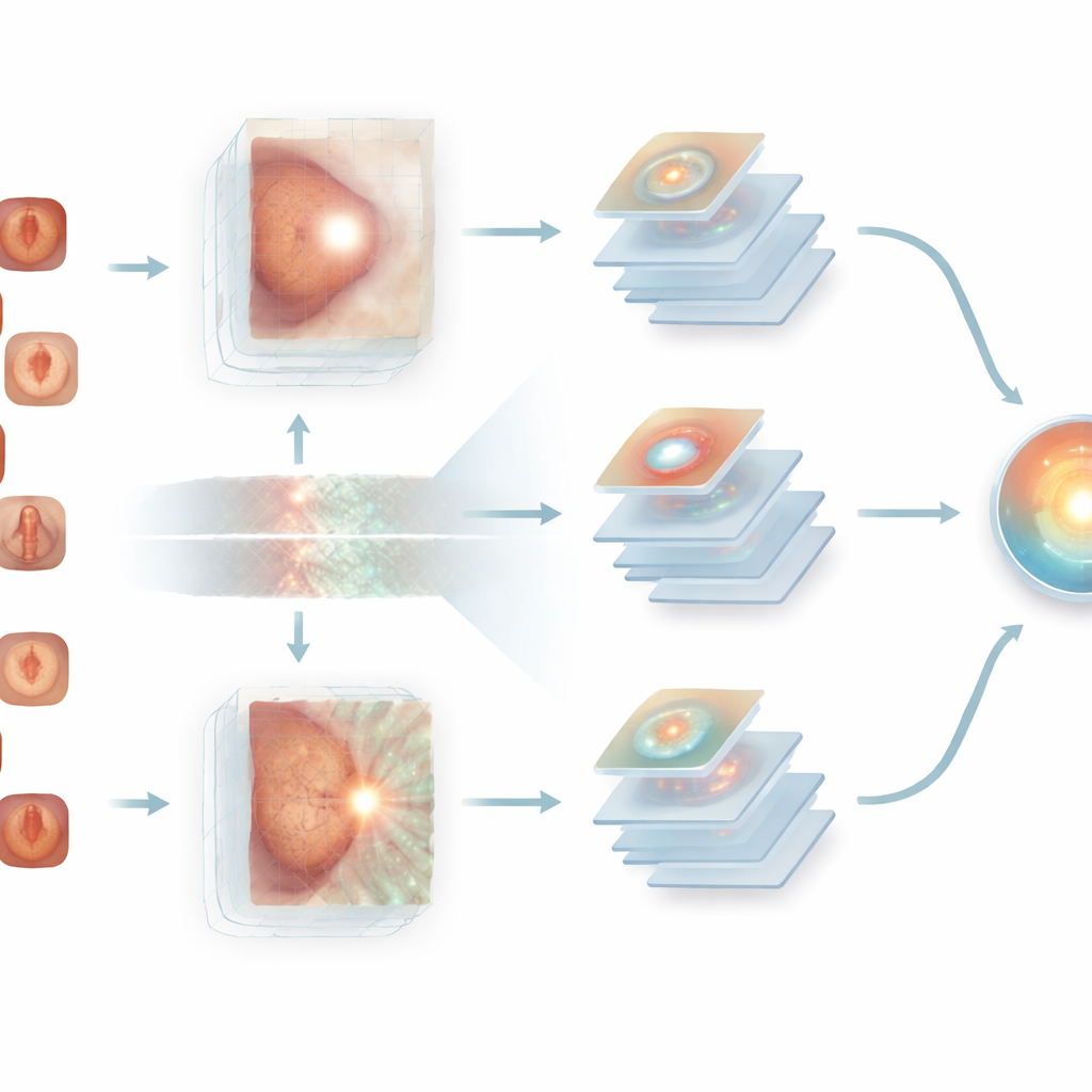
How well does it really work?
The framework is tested on four widely used datasets—ACRIMA, Drishti-Gs, ORIGA, and RIM‑ONE‑DL—covering hundreds of retinal images annotated by experts. Across these collections, the model achieves strikingly high scores: in some cases, it correctly distinguishes glaucomatous from normal eyes in every single test image, with virtually no false alarms or missed cases, and with error levels close to zero. Careful ablation studies show that each component contributes: the hybrid augmentation improves generalization, attention sharpens the focus on critical regions, and the manta‑ray‑based optimizer gives the final boost toward near‑perfect performance.
What this means for future eye care
For a lay reader, the key message is that this research brings us closer to AI tools that can quietly scan routine eye photographs in the background and highlight patients who may need urgent attention before they notice any symptoms. The proposed system does not replace eye doctors, but it can act as a tireless assistant, especially in clinics with limited specialist access. With further testing on broader patient groups and integration into lightweight software or portable devices, such dual‑stream, attention‑guided AI could support earlier, more consistent glaucoma screening and help preserve sight for millions of people worldwide.
Citation: Atia, A., Abdel-kader, H., Abo-Seida, O.M. et al. A hybrid dual-stream CNN framework with dynamic data augmentation and improved Manta Ray Foraging Optimization for robust glaucoma detection. Sci Rep 16, 12701 (2026). https://doi.org/10.1038/s41598-026-45384-6
Keywords: glaucoma screening, retinal imaging, deep learning, medical AI, optic nerve