Clear Sky Science · en
Multi-parametric interferometric reflectance imaging sensor
Why watching molecules stick matters
Many medical tests and new medicines depend on how strongly two molecules stick together—for example, a drug to its target, or a virus to an antibody. Today’s instruments can watch this binding in real time without fluorescent tags, but they often confuse real molecular binding with background changes in the liquid, such as shifts in temperature or salt content. This paper introduces an improved optical sensor that separates true binding from these distractions, making measurements more accurate, simpler, and easier to scale.
Seeing tiny changes with reflected light
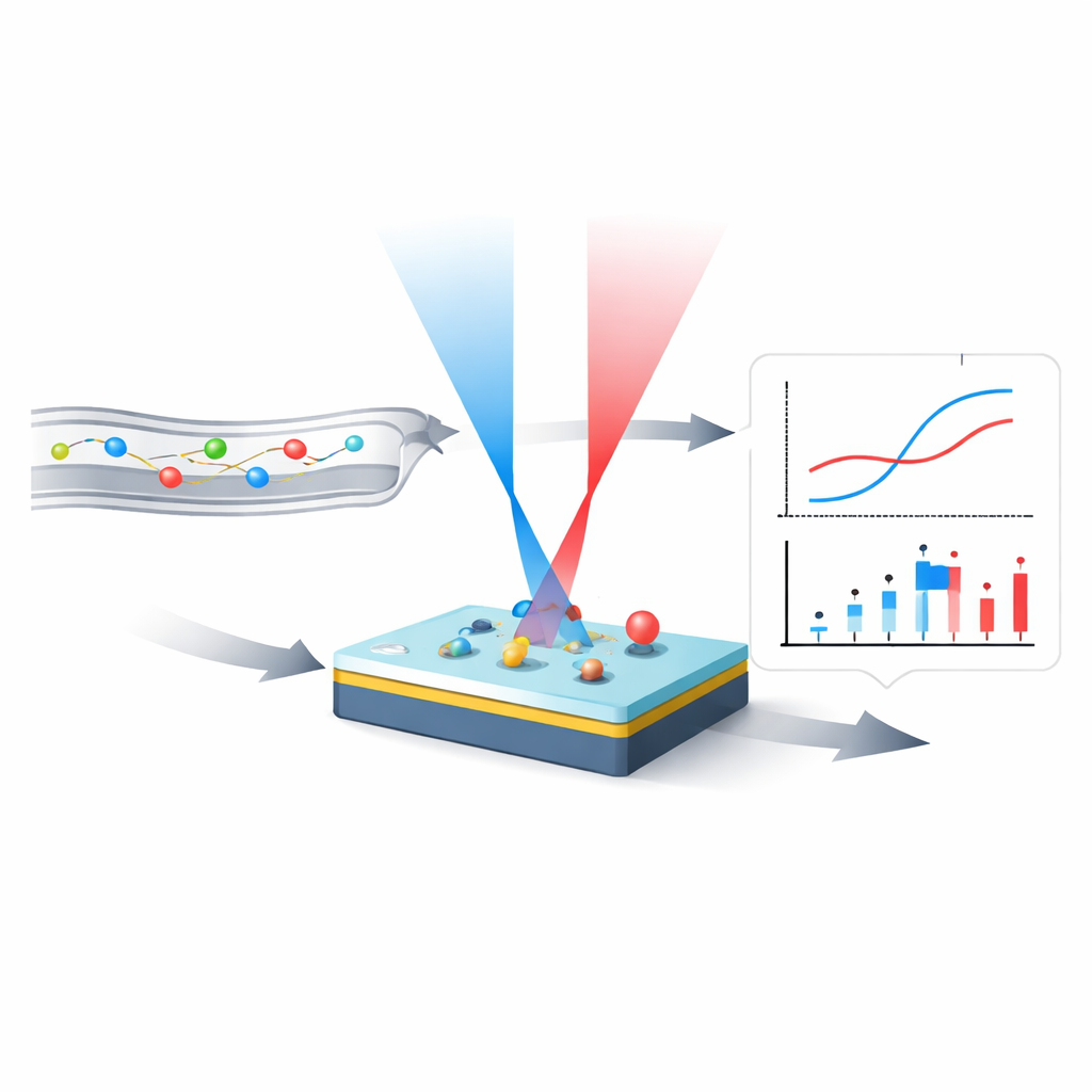
The work builds on an existing technology called an interferometric reflectance imaging sensor, or IRIS. In IRIS, a silicon chip is coated with a very thin transparent layer whose thickness is known. Specific "capture" molecules are attached on top in tiny spots. When target molecules from a flowing solution bind to these spots, they add a minuscule extra thickness. By shining light onto this layered surface and recording how much bounces back, IRIS can translate changes in reflected intensity into changes in apparent thickness, and from there into the amount of material bound at each spot—without needing any molecular labels.
Separating real signals from background shifts
Many other optical sensors rely on an evanescent field that probes just above a metal film. In those systems, any change near the surface—including shifts in temperature, salt, or additives like dimethyl sulfoxide (DMSO)—looks similar to genuine binding, creating what is called the "bulk effect." IRIS is less sensitive to these solution changes, but not completely immune. The authors introduce a multi‑parametric version of IRIS (MP‑IRIS) that reads not only the signal from the spots themselves but also carefully chosen reference areas. By tracking how the response of the surface changes when the liquid composition changes, the system can mathematically remove most of the bulk effect in real time. Experiments in which DMSO concentration was intentionally varied showed that the corrected MP‑IRIS signal reduced bulk error to about 3 picograms per square millimeter—roughly on the order of a few parts per billion of a thin molecular layer, and far below what is typically seen in common commercial instruments.
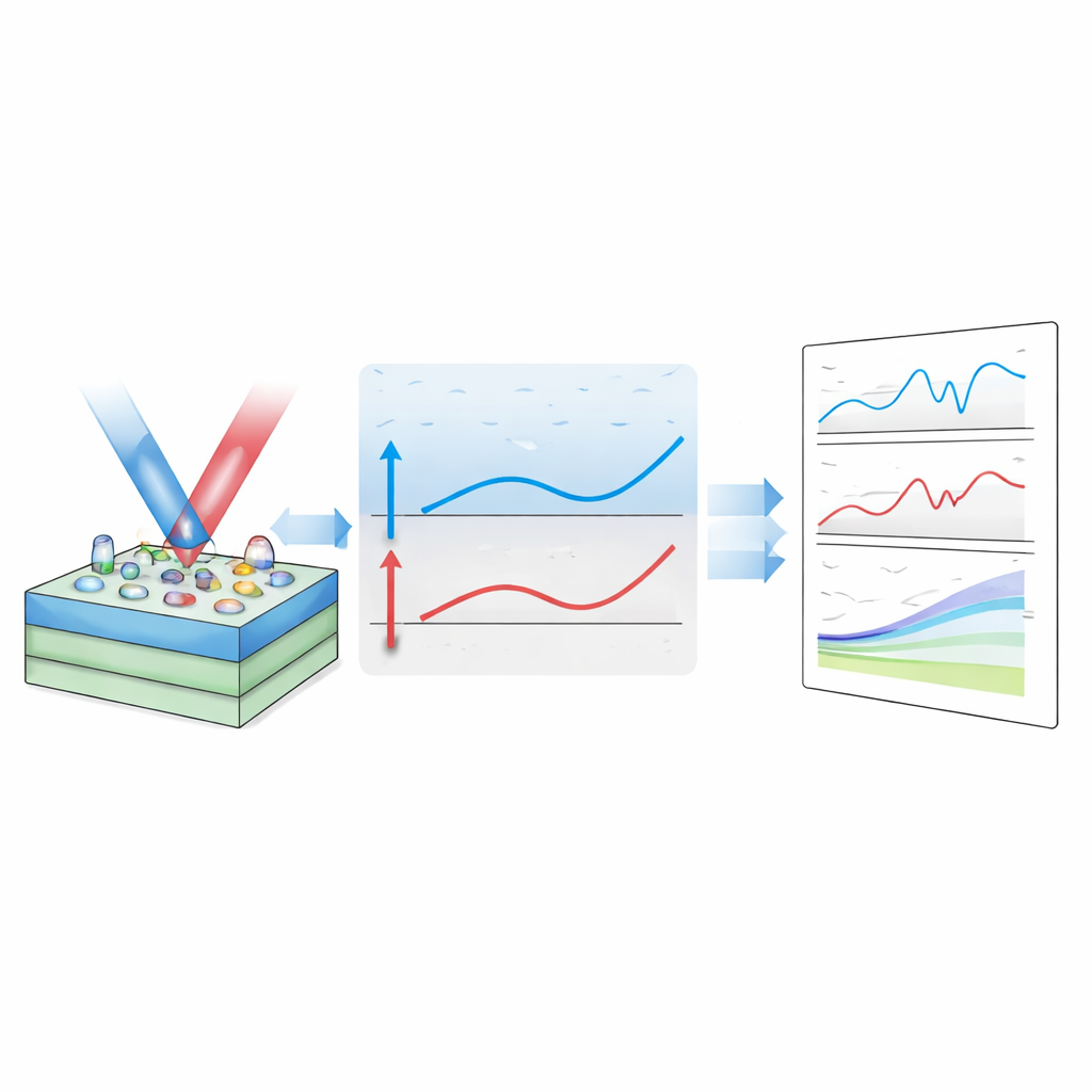
Using two colors of light for robust measurements
While single‑color MP‑IRIS already suppresses background effects, it assumes that the thin layer on the chip has a very precise starting thickness. In practice, small variations arise from chip manufacturing and surface chemistry steps, and these can distort absolute measurements of how much material binds. To overcome this, the authors introduce a second wavelength of light. One color is chosen so that the reflected signal responds strongly to changes in thickness, while the other sits at a point where its reflectance hardly changes with thickness but still reports on the optical properties of the surrounding liquid. By combining readings from these two colors, the system can continuously estimate its own sensitivity, correct for chip‑to‑chip differences, and still remove bulk effects. Tests with deliberate variations in the protein coating showed that two‑color MP‑IRIS kept errors in measured binding below about 10%, even when a simpler approach mis-estimated the same binding by as much as 60%.
Putting the sensor to work with DNA
To demonstrate real biological use, the team ran DNA hybridization experiments. They printed tiny arrays of a protein that captures biotin, then attached short biotin‑tagged DNA strands to some of these spots. When complementary DNA solutions were flowed over the chip, MP‑IRIS recorded, for dozens of spots at once, how the apparent thickness increased as strands found and bound to their partners and then how it changed when the solution without DNA was reintroduced. These tests confirmed that the sensor can follow binding and unbinding in real time, across multiple locations, while correcting for changes in buffer composition and for differences in how heavily each spot was coated.
What this means for future tests
In everyday terms, the new MP‑IRIS design gives scientists a sharper pair of eyes for watching molecules interact. By using smart comparisons between different regions on the chip and between two colors of light, the system largely subtracts out the "background noise" created by the liquid itself and by minor differences between chips. This makes it easier to compare results across experiments and laboratories, and it opens the door to reliable, label‑free tests for small molecules, DNA, proteins, and possibly even viruses, using simpler and more scalable hardware. Further work will explore how the method performs in a wider range of real‑world diagnostic and drug‑screening applications.
Citation: Aslan, M., Snekvik, S., Seymour, E. et al. Multi-parametric interferometric reflectance imaging sensor. Sci Rep 16, 10780 (2026). https://doi.org/10.1038/s41598-026-45282-x
Keywords: label-free biosensing, binding kinetics, optical interferometry, bulk effect correction, DNA hybridization